Review
. 2013 Jun;13(6):438-51.
doi: 10.1038/nri3459.

Immunology of age-related macular degeneration

Affiliations
Review

Immunology of age-related macular degeneration

Jayakrishna Ambati et al. Nat Rev Immunol. 2013 Jun.

Abstract

Age-related macular degeneration (AMD) is a leading cause of blindness in aged individuals. Recent advances have highlighted the essential role of immune processes in the development, progression and treatment of AMD. In this Review we discuss recent discoveries related to the immunological aspects of AMD pathogenesis. We outline the diverse immune cell types, inflammatory activators and pathways that are involved. Finally, we discuss the future of inflammation-directed therapeutics to treat AMD in the growing aged population.


Figures
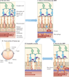
Figure 1
Figure 1. Retinal anatomy in health and disease
The retinal anatomy is composed of several layers (part a); and a cross-section of the human eye (part b) shows focusing of light into the macular area, which is a dense collection of retinal photoreceptors. Normal retinal architecture (part a) is comprised of (from anterior to posterior) a ganglion cell layer, resident retinal microglia, bipolar cells, horizontal cells, the photoreceptor layer, the retinal pigmented epithelium (RPE), the Bruch membrane and a choroidal vascular network. Normal microglia migrate into and out of the subretinal space (as shown by the dashed arrow). Early or intermediate dry age-related macular degeneration (AMD) (part c) is associated with the accumulation of subretinal drusen and microglia and of choroidal macrophages and a thickened Bruch membrane. Geographic atrophy (part d) is the advanced form of dry AMD, which is characterized by confluent regions of RPE and photoreceptor degeneration as well as constriction of choroidal blood vessels. Neovascular AMD (part e) is characterized by the invasion of abnormal, leaky choroidal blood vessels and accompanying macrophages into the retina through breaks in the Bruch membrane, which leads to photoreceptor degeneration.
Figure 2
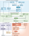
Figure 2. An integrated model of AMD pathogenesis
The cell types, environmental factors and immune pathways that contribute to age-related macular degeneration (AMD) are categorized as either impaired immune-mediated retinal maintenance (part a) or immune-mediated retinal damage (part b). The combination of these factors leads to either geographic atrophy (part c) or neovascular AMD (part d). The most important pathways are highlighted in bold. The earliest known steps in AMD pathogenesis (part a) reflect a reduced capacity to manage the metabolic demands of the retina. The convergence of genetic, environmental and metabolic factors leads to a state in which the accumulation of toxic elements (such as lipid peroxidation by-products, lipofuscin and Alu RNAs) ultimately tips the balance towards immune activation. Once it is replete with unwanted waste material (part b), the retina is the target of inappropriate immune activation. The toxic contents of the retina induce inappropriate activation of diverse immune pathways, including classical and alternative complement pathways, the inflammasome and Toll-like receptor (TLR) signalling. Ultimately, the sustained activation of these pro-inflammatory and damaging pathways leads to advanced AMD. In the case of geographic atrophy (part c), sustained damage to the retinal pigmented epithelium (RPE) leads to the development of degeneration of the RPE, the choriocapillaris and, finally, neural retinal cells. In neovascular AMD (part d), breakdown of the blood–retinal barrier results in immune cell trafficking into the retina, which drives vascular endothelial growth factor A (VEGFA)-dependent neovascularization, causing blindness. 4-HNE, 4-hydroxynonenal; A2E, N-retinyl-N-retinylidene ethanolamine; C1q, complement component 1q; CEP, carboxyethylpyrrole; CX3CR1, CX3C-chemokine receptor 1; CXCL10, CXC-chemokine ligand 10; IL, interleukin; iNKT, invariant natural killer T; MDA, malondialdehyde; TNF, tumour necrosis factor.

Comment in

Similar articles

Cited by

References

    1. The Global Economic Cost of Visual Impairment. AMD Alliance International. 2010 [online], http://www.icoph.org/resources/146/The-Global-Economic-Cost-of-Visual-Im....
    1. Haines JL, et al. Complement factor H variant increases the risk of age-related macular degeneration. Science. 2005;308:419–421. - PubMed
    1. Edwards AO, et al. Complement factor H polymorphism and age-related macular degeneration. Science. 2005;308:421–424. - PubMed
    1. Klein RJ, et al. Complement factor H polymorphism in age-related macular degeneration. Science. 2005;308:385–389. References – are landmark studies showing increased statistical risk of AMD in individuals who have a single CFH polymorphism these studies were the first of their kind for a complex human disease.

    1. Hageman GS, et al. A common haplotype in the complement regulatory gene factor H (HF1/CFH) predisposes individuals to age-related macular degeneration. Proc. Natl Acad. Sci. USA. 2005;102:7227–7232. - PMC - PubMed

Publication types

Substances