. 2013 May 23;4(5):e641.
doi: 10.1038/cddis.2013.165.

Acquired resistance to zoledronic acid and the parallel acquisition of an aggressive phenotype are mediated by p38-MAP kinase activation in prostate cancer cells

Affiliations

Acquired resistance to zoledronic acid and the parallel acquisition of an aggressive phenotype are mediated by p38-MAP kinase activation in prostate cancer cells

M R Milone et al. Cell Death Dis. .

Erratum in

  • Cell Death Dis. 2014;5:e1276

Abstract

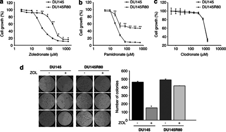
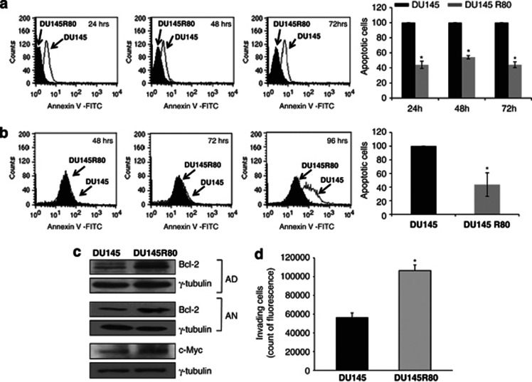
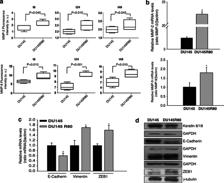
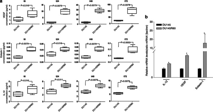
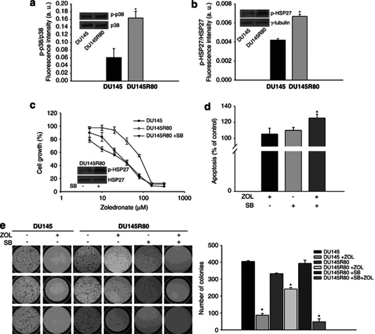
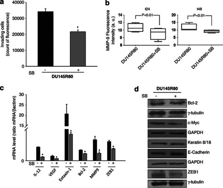
The nitrogen-containing bisphosphonates (N-BP) zoledronic acid (ZOL) inhibits osteoclast-mediated bone resorption, and it is used to prevent skeletal complications from bone metastases. ZOL has also demonstrated anticancer activities in preclinical models and, recently, in cancer patients, highlighting the interest in determining eventual mechanisms of resistance against this agent. In our study, we selected and characterised a resistant subline of prostate cancer (PCa) cells to better understand the mechanisms, by which tumour cells can escape the antitumour effect of ZOL. DU145R80-resistant cells were selected in about 5 months using stepwise increasing concentrations of ZOL from DU145 parental cells. DU145R80 cells showed a resistance index value of 5.5 and cross-resistance to another N-BP, pamidronate, but not to the non-nitrogen containing BP clodronate. Notably, compared with DU145 parental cells, DU145R80 developed resistance to apoptosis and anoikis, as well as overexpressed the anti-apoptotic protein Bcl-2 and oncoprotein c-Myc. Moreover, DU145R80 cells underwent epithelial to mesenchymal transition (EMT) and showed increased expression of the metalloproteases MMP-2/9, as well as increased invading capability. Interestingly, compared with DU145, DU145R80 cells also increased the gene expression and protein secretion of VEGF and the cytokines Eotaxin-1 and IL-12. At the molecular level, DU145R80 cells showed strong activation of the p38-MAPK-dependent survival pathway compared with parental sensitive cells. Moreover, using the p38-inhibitor SB203580, we completely reversed the resistance to ZOL, as well as EMT marker expression and invasion. Furthermore, SB203580 treatment reduced the expression of VEGF, Eotaxin-1, IL-12, MMP-9, Bcl-2 and c-Myc. Thus, for the first time, we demonstrate that the p38-MAPK pathway can be activated under continuous extensive exposure to ZOL in PCa cells and that the p38-MAPK pathway has a critical role in the induction of resistance, as well as in the acquisition of a more aggressive and invasive phenotype.


Figures

Figure 1
Figure 1
Development of ZOL-resistant DU145R80 PCa cell line. DU145 sensitive and DU145R80 resistant cells were treated with increasing concentrations of (a) ZOL, (b) Pamidronate or (c) Clodronate, for 96 h and cell growth assessment was done by sulforhodamine B colorimetric assay (see Materials and Methods). Cell growth is expressed as percentage of control for each time-point. Values are the mean±S.D. from at least three independent experiments performed in quadruplicates (**P⩽0.001 and *P⩽0.05, DU145R80 versus DU145). (d) Soft-agar clonogenic assay was performed on DU145 and DU145R80 cells untreated or treated with ZOL (20 μℳ) for 14 days, in 24-well plates. Colonies >100 μm were scored by a colony counter. Left: images from a representative experiment; right: values expressed as number of colonies are means±S.D. from at least two independent experiments performed in triplicates. Statistical analysis demonstrated significant differences only in DU145 cells (*P<0.001, ZOL versus untreated)
Figure 2
Figure 2
Resistance to apoptosis and anoikis and increased invasiveness of DU145R80 compared with DU145 cells. Apoptosis evaluated by AnnexinV binding and cytofluorimetric analysis in DU145 compared with DU145R80 cells, in adherent conditions (a), and non-adherent conditions (anoikis) (b), for the indicated time points (see Materials and Methods). Left: representative experiment; right: values of DU145R80 apoptotic cells expressed as per cent of DU145 parental cells (100%). Values are means±S.D. of three independent experiments. Statistical analysis demonstrated significant differences for all time points in adherent conditions (*P=0.007, 24 h; P=0.002, 48 h; P=0.005, 72 h) and at 96 h in non-adherent conditions (*P=0.031). (c) Western blot analysis of Bcl-2 in adherent (AD) and in anoikis conditions (AN) and of c-Myc. Cell lysates were obtained after 48 h of cell culture. γ-tubulin was used as protein loading control. (d) Invasion capability of DU145 and DU145R80 cells was evaluated as the ability to invade matrigel coated chambers (see Materials and Methods). Values are means±S.D. from three independent experiments performed in duplicates and statistical analysis is reported (*P=0.023, DU145R80 versus DU145 cells)
Figure 3
Figure 3
Increased expression of MMPs and EMT features in DU145R80 compared with DU145 cells. (a) MMP-2 and MMP-9 concentrations were evaluated in the culture media from DU145 and DU145R80 cells at the indicated time points after seeding by a multiplex ELISA-based immunoassay assay, and were plotted with box-and-whisker graphs. The boxes extend from the 25th to the 75th percentile and the line in the middle is the median. The error bars extend down to the lowest value and up to the highest. (b) MMP-2 and MMP-9 mRNA expression was evaluated by RT-PCR after 24 and 6 h of cell culture, respectively. The data are representative of at least three independent experiments, include the means±S.D. of technical triplicates and reported statistical analysis of DU145R80 versus DU145 cells (*P<0.001, MMP-2; P=0.045, MMP-9). (c) E-cadherin, Vimentin and ZEB1 mRNA expression was evaluated by RT-PCR after 24 h of cell culture. The data are representative of at least three independent experiments, include the means±S.D. of technical triplicates and reported statistical analysis of DU145R80 versus DU145 cells (*P=0.02, E-cadherin; P=0.007, Vimentin; P=0.005, ZEB1). (d) Western blot analysis of Keratin 8/18, E-cadherin, Vimentin and ZEB1 proteins expression evaluated after 48 h of cell culture. GAPDH and γ-tubulin were used as protein loading control
Figure 4
Figure 4
Increased expression of VEGF, Eotaxin-1 and IL-12 in DU145R80 compared with DU145 cells. (a) VEGF, Eotaxin-1 and IL-12 concentrations were evaluated in the culture media from DU145 and DU145R80 cells at the indicated time points after seeding by a multiplex ELISA-based immunoassay assay, and were plotted with box-and-whisker graphs. The boxes extend from the 25th to the 75th percentile, and the line in the middle is the median. The error bars extend down to the lowest value and up to the highest. (b) IL-12, VEGF and Eotaxin-1 mRNA expression was evaluated by RT-PCR after 6 h of cell culture. The data are representative of at least three independent experiments, include the means±S.D. of technical triplicates and reported statistical analysis of DU145R80 versus DU145 cells (*P<0.001, IL-12 and VEGF; P=0.014, Eotaxin-1)
Figure 5
Figure 5
P38-MAPK activation was involved in the resistance to ZOL of DU145R80 cells. (a) Analysis of p38-MAPK and HSP27 activation evaluated by a phosphoprotein ELISA-based immunoassay (see Materials and Methods) (a and b) and by western blot (a and b inset panels). Values from phosphoprotein assay are means±S.D. of two independent experiments performed in triplicates and statistical analysis of DU145R80 versus DU145 cells is reported (*P=0.037, p38-MAPK; P=0.005, HSP27). (c) DU145R80 cells were treated for 96 h with increasing concentrations of ZOL alone or after 24 h of pretreatment with SB 30 μℳ and compared with DU145 treated with ZOL alone. Cell growth expressed as percentage of control was assessed by sulforhodamine B colorimetric assay (see Materials and Methods) and each point is the mean±S.D. of three independent experiments. Expression of p-HSP27 and HSP-27 in DU145R80 cells untreated or treated with SB 30 μℳ for 24 h were evaluated by western blot (inset panel). (d) Apoptosis evaluated by AnnexinV binding and cytofluorimetric analysis in DU145R80 cells untreated or treated with ZOL 20 μℳ alone or in combination with SB 30 μℳ for 48 h. Values of apoptotic cells are means±S.D. of three independent experiments and were expressed as per cent of untreated cells (100%). Statistical analysis demonstrated significant differences only in ZOL+SB combination versus untreated cells (*P=0.026). (e) Soft-agar clonogenic assay was performed on DU145 and DU145R80 cells untreated or treated with ZOL alone (20 μℳ) or in combination with SB (30 μℳ) for 21 days, in 24-well plates. Colonies >100 μm were scored by a colony counter. Left: images from a representative experiment; right: values expressed as number of colonies are means±S.D. from at least two independent experiments performed in triplicates. Statistical analysis results are reported (*P<0.001, ZOL versus untreated cells in DU145; P=0.005, ZOL versus untreated cells in DU145R80; P=0.002, P=0.005, P=0.003, ZOL+SB combination versus untreated, versus ZOL or versus SB-treated cells, respectively, in DU145R80)
Figure 6
Figure 6
P38-MAPK activation regulated invasion, EMT and the expression of Bcl-2, c-Myc, MMP9 and cytokines in DU145R80. (a) Invasion capability of DU145R80 cells untreated or treated with SB (30 μℳ) for 24 h was evaluated as the ability to invade matrigel coated chambers (see Materials and Methods). Values are means±S.D. from three independent experiments performed in duplicates and statistical analysis is reported (*P<0.001). (b) MMP-9 concentration was evaluated in the culture media of DU145R80 cells untreated or treated with SB (30 μℳ) for the indicated time points after seeding, by a multiplex ELISA-based immunoassay assay, and were plotted with box-and-whisker graphs. The boxes extend from the 25th to the 75th percentile and the line in the middle is the median. The error bars extend down to the lowest value and up to the highest. (c) IL-12, VEGF, Eotaxin-1, Bcl-2, MMP9 and ZEB1 mRNA expression was evaluated by RT-PCR in DU145R80 cells untreated or treated with SB (30 μℳ) for 6 h. Values are means±S.D. from three independent experiments performed in duplicates and statistical analysis is reported (*P<0.01). (d) Western blot analysis of Bcl2, c-Myc, Keratin 8/18, E-cadherin and ZEB1 proteins expression evaluated in DU145R80 cells untreated or treated with SB (30 μℳ) for 72 h of cell culture. GAPDH and γ-tubulin were used as protein loading control
Figure 7
Figure 7
Proposed model for the mechanisms underlying the acquired resistance to ZOL and the parallel acquisition of an aggressive phenotype mediated by p38-MAP Kinase activation

Similar articles

Cited by

References

    1. Haas GP, Delongchamps N, Brawley OW, Wang CY, de la Roza G. The worldwide epidemiology of prostate cancer: perspectives from autopsy studies. Can J Urol. 2008;15:3866–3871. - PMC - PubMed
    1. Mahon KL, Henshall SM, Sutherland RL, Horvath LG. Pathways of chemotherapy resistance in castration-resistant prostate cancer. Endocr Relat Cancer. 2011;18:R103–R123. - PubMed
    1. Coleman R, Gnant M, Morgan G, Clezardin P. Effects of bone-targeted agents on cancer progression and mortality. J Natl Cancer Inst. 2012;104:1059–1067. - PubMed
    1. Saylor PJ, Lee RJ, Smith MR. Emerging therapies to prevent skeletal morbidity in men with prostate cancer. J Clin Oncol. 2011;29:3705–3714. - PMC - PubMed
    1. Santini D, Caraglia M, Vincenzi B, Holen I, Scarpa S, Budillon A, et al. Mechanisms of disease: preclinical reports of antineoplastic synergistic action of bisphosphonates. Nat Clin Pract Oncol. 2006;3:325–338. - PubMed

Publication types

MeSH terms

LinkOut - more resources