. 2013 Sep;8(9):1515-23.
doi: 10.2215/CJN.10491012. Epub 2013 May 23.

Renal amyloidosis: origin and clinicopathologic correlations of 474 recent cases

Affiliations

Renal amyloidosis: origin and clinicopathologic correlations of 474 recent cases

Samar M Said et al. Clin J Am Soc Nephrol. 2013 Sep.

Abstract

Background and objectives: The kidney is the organ most commonly involved in systemic amyloidosis. This study reports the largest clinicopathologic series of renal amyloidosis.

Design, setting, participants, & measurements: This study provides characteristics of 474 renal amyloidosis cases evaluated at the Mayo Clinic Renal Pathology Laboratory from 2007 to 2011, including age, sex, serum creatinine, proteinuria, type of amyloid, and tissue distribution according to type.

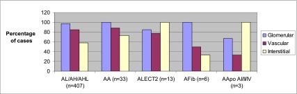
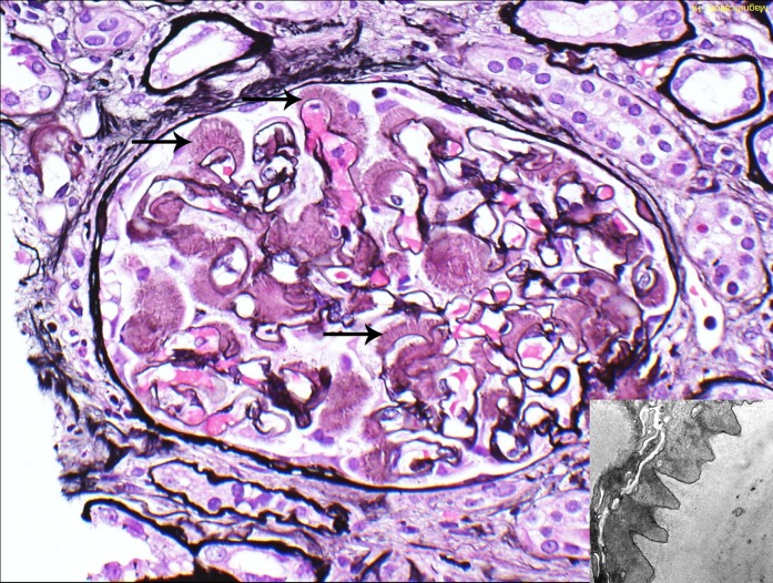
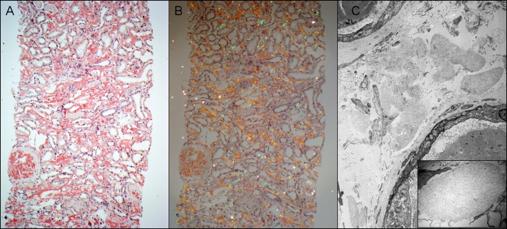
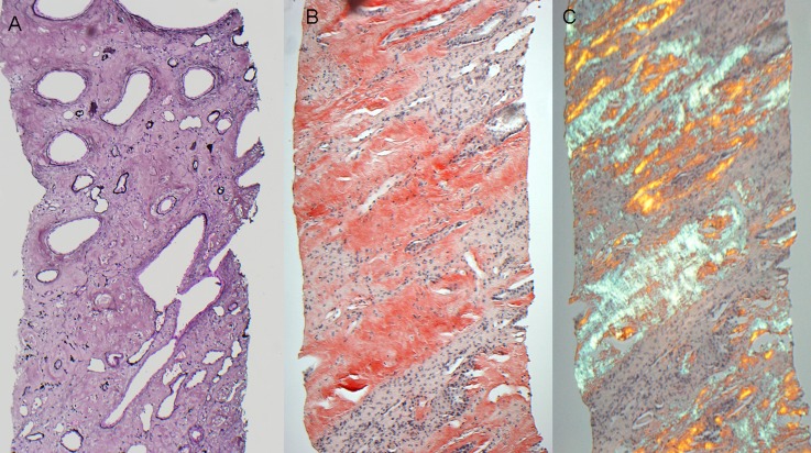
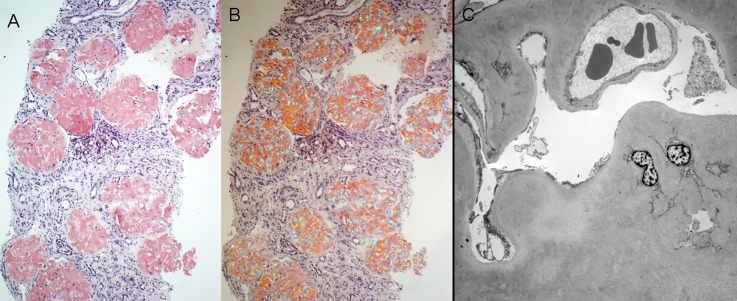
Results: The type of amyloid was Ig amyloidosis in 407 patients (85.9%), AA amyloidosis in 33 (7.0%), leukocyte chemotactic factor 2 amyloidosis in 13 (2.7%), fibrinogen A α chain amyloidosis in 6 (1.3%), Apo AI, Apo AII, or Apo AIV amyloidosis in 3 (0.6%), combined AA amyloidosis/Ig heavy and light chain amyloidosis in 1 (0.2%), and unclassified in 11 (2.3%). Laser microdissection/mass spectrometry, performed in 147 cases, was needed to determine the origin of amyloid in 74 of the 474 cases (16%), whereas immunofluorescence failed to diagnose 28 of 384 light chain amyloidosis cases (7.3%). Leukocyte chemotactic factor 2 amyloidosis and Apo AI, Apo AII, or Apo AIV amyloidosis were characterized by diffuse interstitial deposition, whereas fibrinogen A α chain amyloidosis showed obliterative glomerular involvement. Compared with other types, Ig amyloidosis was associated with lower serum creatinine, higher degree of proteinuria, and amyloid spicules.

Conclusions: In the authors' experience, the vast majority of renal amyloidosis cases are Ig derived. The newly identified leukocyte chemotactic factor 2 amyloidosis form was the most common of the rarer causes of renal amyloidosis. With the advent of laser microdissection/mass spectrometry for amyloid typing, the origin of renal amyloidosis can be determined in >97% of cases.


Figures

Figure 1.
Figure 1.
Distribution of renal amyloid deposits according to type. Glomerular involvement: ALECT2/AApo AI/AII/AIV versus AL/AH/AHL/AA/AFib, P=0.01. Vascular involvement: ALECT2/AFib/AApo AI/AII/AIV versus AL/AH/AHL/AA, P=0.01; AFib/AApo AI/AII/AIV versus AL/AH/AHL/AA/ALECT2, P=0.01. Interstitial involvement: ALECT2/AApo AI/AII/AIV versus AL/AH/AHL/AA/AFib, P<0.001. AL/AH/AHL, light chain/heavy chain/heavy and light chain amyloidosis; AA, AA amyloidosis; ALECT2, leukocyte chemotactic factor 2 amyloidosis; AFib, fibrinogen A α-chain amyloidosis; AApo AI/AII/AIV, Apo AI, Apo AII, or Apo AIV amyloidosis.
Figure 2.
Figure 2.
Glomerular amyloid spicules. Glomerular amyloid spicules (arrows) result from parallel alignment of amyloid fibrils in the subepithelial zone perpendicular to the glomerular basement membrane. They were more common in AL/AH/AHL than other types of amyloidosis (Jones methenamine silver). The small image shows amyloid spicules by electron microscopy. AL/AH/AHL, light chain/heavy chain/heavy and light chain amyloidosis. Original magnification, ×400 in image; ×23,000 in inset.
Figure 3.
Figure 3.
ALECT2. (A) This case of ALECT2 exhibits diffuse cortical interstitial amyloid deposition. The glomerulus on the left is also globally involved (Congo red stain). (B) The Congo red-positive amyloid deposits show apple-green birefringence when viewed under polarized light. (C) Electron microscopy shows several interstitial aggregates of amyloid deposits. The small image is a higher magnification showing the fibrillar nature of deposits. ALECT2, leukocyte chemotactic factor 2 amyloidosis. Original magnification, ×100 in A and B; ×5800 in C; ×46,000 in C inset.
Figure 4.
Figure 4.
AApo AI/AII/AIV. (A) This case of AApo AI shows diffuse medullary interstitial amyloid deposits, which stain silver negative. (B) This case of AApo AIV reveals diffuse medullary interstitial amyloid deposits that stain strongly Congo red positive. (C) The Congo red stained sections from the case shown in B exhibit apple-green birefringence when viewed under polarized light. AApo AI/AII/AIV, Apo AI, Apo AII, or Apo AIV amyloidosis. Original magnification, ×100.
Figure 5.
Figure 5.
AFib. (A) There is diffuse obliterative glomerular involvement by amyloidosis in this case of AFib (Congo red stain). (B) The Congo red-stained sections from the case shown in A exhibit apple-green birefringence when viewed under polarized light. (C) Electron microscopy shows marked mesangial and glomerular capillary wall amyloid deposition. Despite the subtotal obliteration of glomerular capillary lumina by amyloid deposits, no amyloid spicules are seen. AFib, fibrinogen A α chain amyloidosis. Original magnification, ×100 in A and B; ×1900 in C.

References

    1. Eulitz M, Weiss DT, Solomon A: Immunoglobulin heavy-chain-associated amyloidosis. Proc Natl Acad Sci U S A 87: 6542–6546, 1990 - PMC - PubMed
    1. Benson MD, Liepnieks J, Uemichi T, Wheeler G, Correa R: Hereditary renal amyloidosis associated with a mutant fibrinogen alpha-chain. Nat Genet 3: 252–255, 1993 - PubMed
    1. Soutar AK, Hawkins PN, Vigushin DM, Tennent GA, Booth SE, Hutton T, Nguyen O, Totty NF, Feest TG, Hsuan JJ, Pepys MB: Apolipoprotein AI mutation Arg-60 causes autosomal dominant amyloidosis. Proc Natl Acad Sci U S A 89: 7389–7393, 1992 - PMC - PubMed
    1. Benson MD, Liepnieks JJ, Yazaki M, Yamashita T, Hamidi Asl K, Guenther B, Kluve-Beckerman B: A new human hereditary amyloidosis: The result of a stop-codon mutation in the apolipoprotein AII gene. Genomics 72: 272–277, 2001 - PubMed
    1. Sethi S, Theis JD, Shiller SM, Nast CC, Harrison D, Rennke HG, Vrana JA, Dogan A: Medullary amyloidosis associated with apolipoprotein A-IV deposition. Kidney Int 81: 201–206, 2012 - PubMed

Publication types

MeSH terms