

Antioxidants inhibit SAA formation and pro-inflammatory cytokine release in a human cell model of alkaptonuria
- PMID: 23704321
- PMCID: PMC3741479
- DOI: 10.1093/rheumatology/ket185
Antioxidants inhibit SAA formation and pro-inflammatory cytokine release in a human cell model of alkaptonuria
Abstract
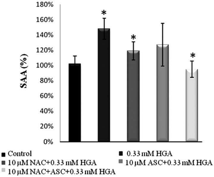
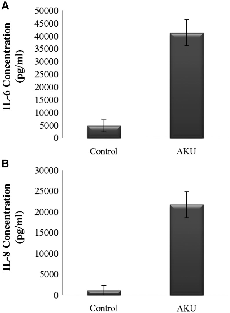
Objective: Alkaptonuria (AKU) is an ultra-rare autosomal recessive disease that currently lacks an appropriate therapy. Recently we provided experimental evidence that AKU is a secondary serum amyloid A (SAA)-based amyloidosis. The aim of the present work was to evaluate the use of antioxidants to inhibit SAA amyloid and pro-inflammatory cytokine release in AKU.
Methods: We adopted a human chondrocytic cell AKU model to evaluate the anti-amyloid capacity of a set of antioxidants that had previously been shown to counteract ochronosis in a serum AKU model. Amyloid presence was evaluated by Congo red staining. Homogentisic acid-induced SAA production and pro-inflammatory cytokine release (overexpressed in AKU patients) were evaluated by ELISA and multiplex systems, respectively. Lipid peroxidation was evaluated by means of a fluorescence-based assay.
Results: Our AKU model allowed us to prove the efficacy of ascorbic acid combined with N-acetylcysteine, taurine, phytic acid and lipoic acid in significantly inhibiting SAA production, pro-inflammatory cytokine release and membrane lipid peroxidation.
Conclusion: All the tested antioxidant compounds were able to reduce the production of amyloid and may be the basis for establishing new therapies for AKU amyloidosis.
Keywords: N-acetylcysteine; ascorbic acid; chondrocyte; homogentisic acid; inflammation; lipid peroxidation; lipoic acid; phytic acid; serum amyloid A; taurine.
Figures


References
-
- Hegedus ZL. The probable involvement of soluble and deposited melanins, their intermediates and the reactive oxygen side-products in human diseases and aging. Toxicology. 2000;145:85–101. - PubMed
-
- Tinti L, Taylor AM, Santucci A, et al. Development of an in vitro model to investigate joint ochronosis in alkaptonuria. Rheumatology. 2010;50:271–7. - PubMed
-
- Braconi D, Laschi M, Amato L, et al. Evaluation of anti-oxidant treatments in an in vitro model of alkaptonuric ochronosis. Rheumatology. 2010;49:1975–83. - PubMed
-
- Tinti L, Spreafico A, Braconi D, et al. Evaluation of antioxidant drugs for the treatment of ochronotic alkaptonuria in an in vitro human cell model. J Cell Physiol. 2010;225:84–91. - PubMed
Publication types
MeSH terms
Substances
Grants and funding
LinkOut - more resources
Full Text Sources
Other Literature Sources
Medical
