

Extramedullary haematopoiesis in axillary lymph nodes following neoadjuvant chemotherapy for locally advanced breast cancer
- PMID: 23704429
- PMCID: PMC3669812
- DOI: 10.1136/bcr-2013-008943
Extramedullary haematopoiesis in axillary lymph nodes following neoadjuvant chemotherapy for locally advanced breast cancer
Abstract
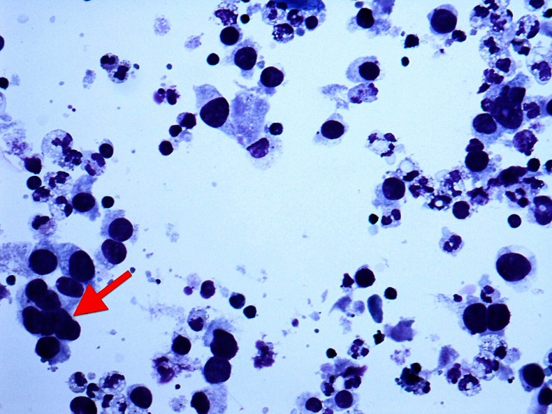
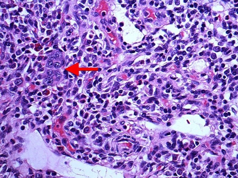
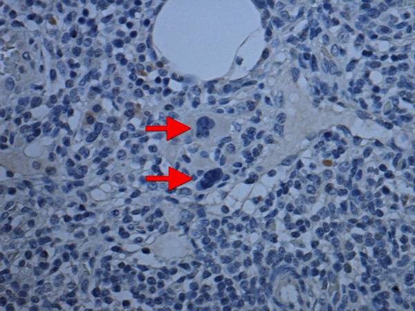
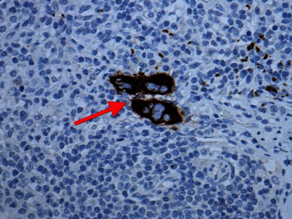
We report the case of a 53-year-old lady who presented with a lump in her left breast. Her initial investigations demonstrated a grade III invasive ductal carcinoma of the breast that was tethered to the pectoralis major; imaging and cytology also revealed metastatic nodes in the left axilla. After undergoing neoadjuvant chemotherapy with evidence of clinical and radiological tumour response, a wire-guided wide local excision and axillary node clearance was performed. When a histological analysis of the specimen was performed, there was no evidence of a viable metastatic tumour in the axillary lymph nodes, but there were several areas of extramedullary haematopoiesis. There are only two other reports in the literature of this finding. This could represent a potential source of false-positive diagnosis of axillary metastasis from breast cancer. It would be prudent to consider biopsy prior to clearance if there are megakaryocytes in axillary node cytology.
Figures
References
-
- Millar E, Inder S, Lynch J. Extramedullary haematopoiesis in axillary lymph nodes following neoadjuvant chemotherapy for locally advanced breast cancer—a potential diagnostic pitfall. Histopathology 2009;2013:622–33 - PubMed
-
- Zafar N. Megakaryocytes in sentinel lymph node—a potential source for diagnostic error. Breast J 2007;2013:308–9 - PubMed
-
- National Institute for Health and Clinical Excellence Guidance on cancer services: improving outcomes in breast cancer. London, UK: National Institute for Clinical Excellence; 2002
-
- D'Souza A, Jaiyesimi I, Trainor L, et al. Granulocyte colony-stimulating factor administration: adverse events. Transfus Med Rev 2008;2013:280–90 - PubMed
-
- Smith I, Heys S, Hutcheon A, et al. Neoadjuvant chemotherapy in breast cancer: significantly enhanced response with docetaxel. J Clin Oncol 2002;2013:1456–66 - PubMed
Publication types
MeSH terms
Substances
LinkOut - more resources
Full Text Sources
Other Literature Sources
Medical