

Porphyria: varied ocular manifestations and management
- PMID: 23704443
- PMCID: PMC3669952
- DOI: 10.1136/bcr-2013-009496
Porphyria: varied ocular manifestations and management
Abstract
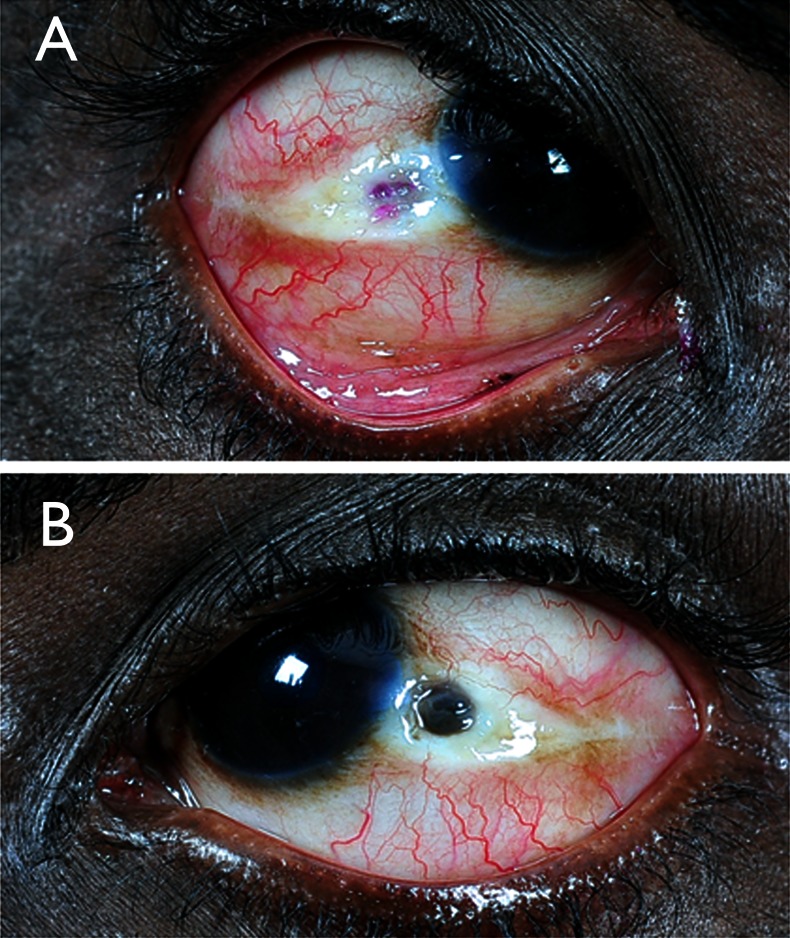
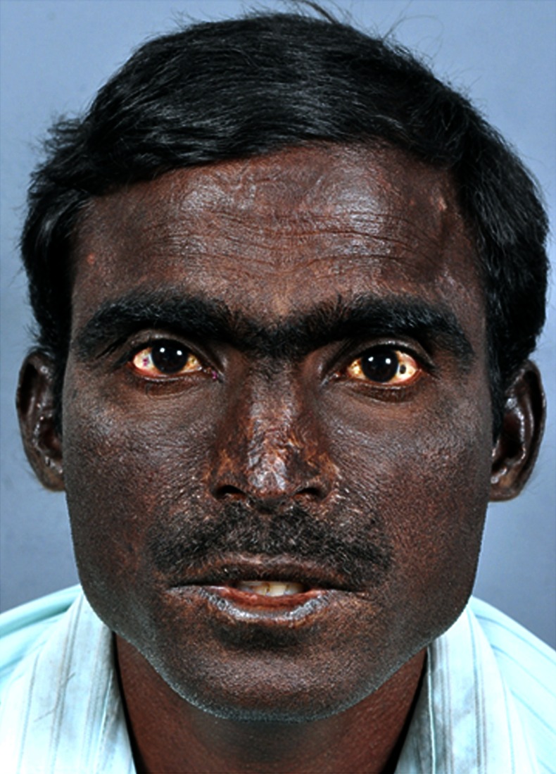
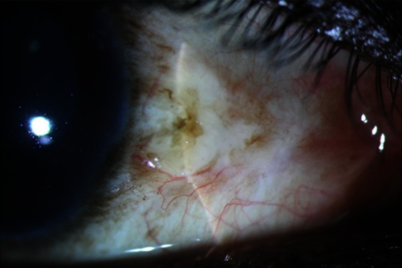
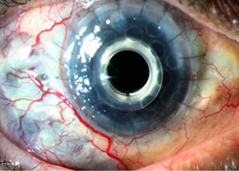
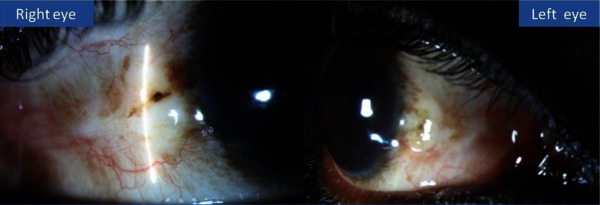
On review of past 10 years medical records, we could find four typical cases of porphyria with rare ocular manifestations. Cases 1, 2 and 4 have presented with features suggestive of acute scleritis. Based on clinical, biochemical and dermatological evaluation, all these three cases were diagnosed to have congenital erythropoietic porphyria. Case 1 was initially managed with scleral patch graft which on subsequent melt was managed with double layered amniotic membrane grafting along with conjunctival advancement and lateral paramedian tarsorrhaphy in both the eyes. Cases 2 and 4 were managed conservatively with artificial tear drops and general protective measures. Case 3 was presented with multiple failed grafts due to repeated ulceration and infection. Owing to multiple failed grafts, Boston keratoprosthesis was done and the patient is doing well with stable kertaoprosthesis at the last follow-up visit.
Figures
References
-
- Venkatesh P, Garg SP, Kumaran E, et al. Congenital porphyria with necrotizing Scleritis in a 9-year-old child. Clin Exp Ophthalmol 2000;2013:314–18 - PubMed
-
- Sober AJ, Grove AS, Muhlbauer JE. Cicatricial ectropion and lacrimal obstruction associated with the sclerodermoid variant of porphyria cutanea tarda. Am J Ophthalmol 1981;2013:396–400 - PubMed
-
- Veenashree MP, Sangwan VS, Vemuganti GK, et al. Acute scleritis as a manifestation of congenital erythropoietic porphyria. Cornea 2012;2013:530–1 - PubMed
Publication types
MeSH terms
LinkOut - more resources
Full Text Sources
Other Literature Sources
Medical
Miscellaneous