

Lateral palatal flap approach to the nasopharynx and parapharyngeal space for transoral robotic surgery: a cadaveric study
- PMID: 23704859
- PMCID: PMC3657084
- DOI: 10.1007/s11701-012-0351-6
Lateral palatal flap approach to the nasopharynx and parapharyngeal space for transoral robotic surgery: a cadaveric study
Abstract
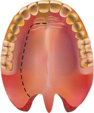
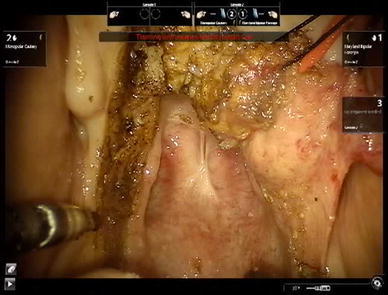
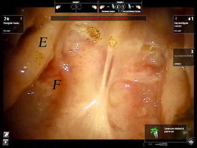
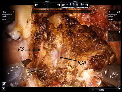
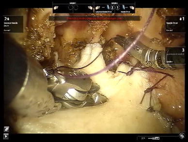
The da Vinci surgical robot has been used for minimally invasive surgery of the head and neck region including resection of tumors in the nasopharynx. Access to and vision of the nasopharynx with the robot are difficult. A pure transoral approach and midline palatal split approach have been described. The disadvantage of these approaches is the limited lateral access to the parapharyngeal space. The objective of this study was to investigate the feasibility of accessing the nasopharynx and parapharyngeal space with a lateral palatal flap. Two complete nasopharyngectomies with resection of the parapharyngeal space and exposure of the internal carotid artery and branches of the mandibular nerves were performed on two fresh cadavers with the da Vinci surgical robot. The set up of the robot, the surgical procedure of elevating the lateral palatal flap, and robotic resection of the nasopharynx and parapharyngeal space are described.
Keywords: Nasopharyngectomy; Recurrent nasopharyngeal cancer; Robotic surgery.
Figures

References
-
- Dallan I, Castelnuovo P, Montevecchi F, Battaglia P, Cerchiai N, Seccia V, Vicini C (2011) Combined transoral transnasal robotic-assisted nasopharyngectomy: a cadaveric feasibility study. Eur Arch Otorhinolaryngol. doi:10.1007/s00405-011-1550-x - PubMed
-
- Wei WI, Ho W-K (2010) Transoral Robotic Resection of Recurrent Nasopharyngeal Carcinoma. The Laryngoscope. doi:10.1002/lary.21059 - PubMed
-
- Yin Tsang RK, Ho WK, Wei WI (2011) Combined transnasal endoscopic and transoral robotic resection of recurrent nasopharyngeal carcinoma. Head Neck. doi:10.1002/hed.21731 - PubMed
LinkOut - more resources
Full Text Sources
