. 2013 May 21;8(5):e62642.
doi: 10.1371/journal.pone.0062642. Print 2013.

Interaction between IGF-IR and ER induced by E2 and IGF-I

Affiliations

Interaction between IGF-IR and ER induced by E2 and IGF-I

Zhenghong Yu et al. PLoS One. .

Abstract

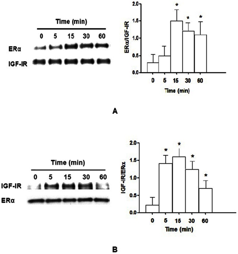
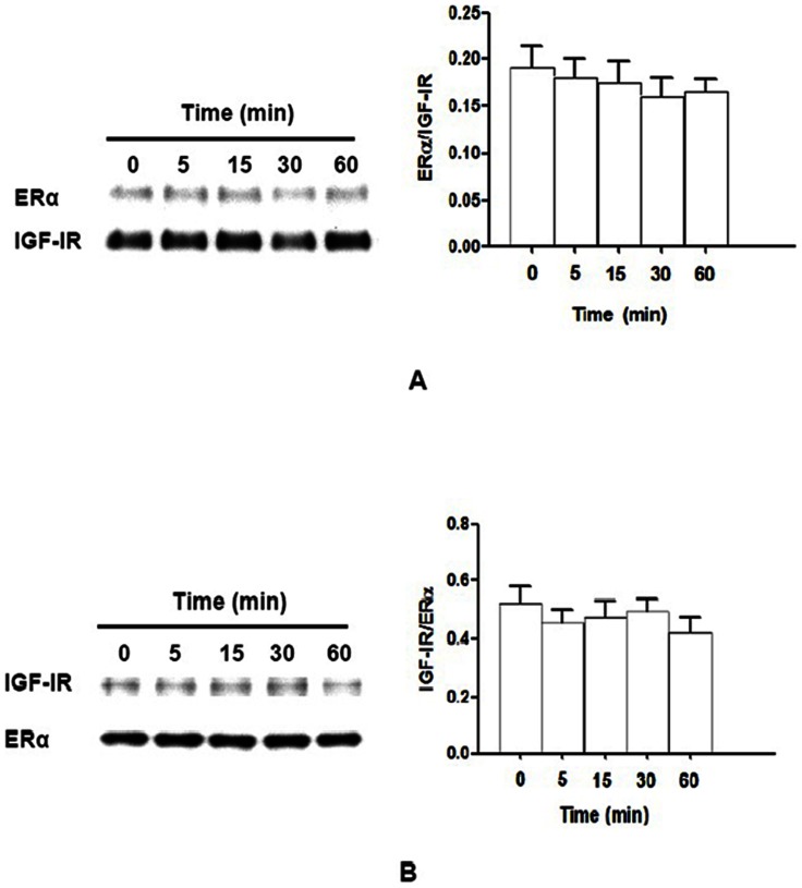
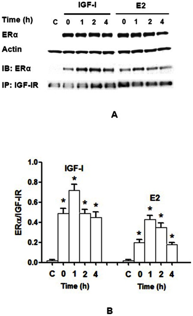
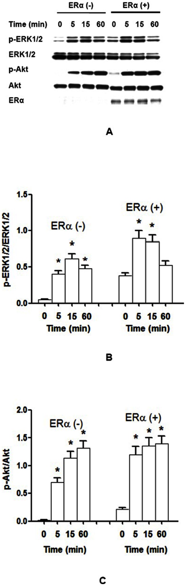
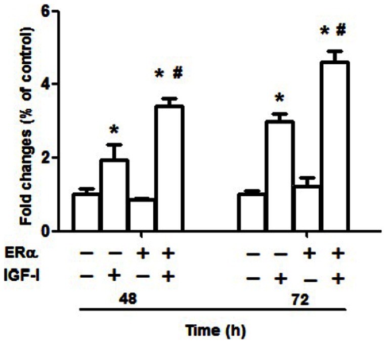
Estrogen receptor (ER) is a nuclear receptor and the insulin-like growth factor-I (IGF-I) receptor (IGF-IR) is a transmembrane tyrosine kinase receptor. Estrogen and IGF-I are known to have synergistic effects on the growth of breast cancer cells. Recently, non-nuclear effects of ER have been under investigation. To study the mechanism involved in this process, we have used MCF-7 breast cancer cell lines transfected with IGF-IR anti-sense cDNA (SX13, MCF-7(SX13)) that resulted in 50% reduction of IGF-IR. In MCF-7 cells, estradiol (E2) and IGF-I induced the rapid association of ER to IGF-IR, however, the interaction was abrogated in MCF-7(SX13) cells. In addition, NWTB3 cells (NIH3T3 cells overexpressing IGF-IR) were transiently transfected with ERα, the ER-IGF-IR interaction was induced by both E2 and IGF-I. Moreover, ERα regulated the IGF-I signaling pathways through phosphorylation of ERK1/2 and Akt and the interaction of ER-IGF-IR potentiated the cell growth. Finally, E2 and IGF-I stimulated translocation of ER from the nucleus to the cytoplasm. Taken together, these findings reveal that the interaction of the ER and IGF-IR is important for the non-genomic effects of ER.


Conflict of interest statement

Competing Interests: The authors have declared that no competing interests exist.

Figures

Figure 1
Figure 1. Binding of IGF-IR to ERα is stimulated by IGF-I in MCF-7 cells.
MCF-7 cells were stimulated with 10 nM IGF-I for 0, 5, 15, 30, or 60 min. Protein lysates were subjected to immunoprecipitation with IGF-IR antibody and subsequently immunoblotted with ERα antibody (A) or immunoprecipitation with ERα antibody and subsequently immunoblotted with IGF-IR antibody (B). The ratio of ERα and IGF-IR rapidly increased from 0 to 15 min, and reached peak at 15 min and gradually decreased from 30 to 60 min (A). Also, the ratio of IGF-IR and ERα rapidly increased from 0 to 15 min, and reached peak at 15 min and gradually decreased from 30 to 60 min (B). N = 3, *, P<0.05 as compared to 0 min.
Figure 2
Figure 2. Binding IGF-IR to ERα is abrogated by IGF-I in MCF-7SX13 cells.
MCF-7SX13 cells were stimulated with 10 nM IGF-I for 0–60 min. Protein lysates were subjected to immunoprecipitation with IGF-IR (A) or ERα (B) antibodies with subsequent immunoblotting of the precipitated fraction with ERα antibody (A) or IGF-IR antibody (B), respectively. The ratios of ERα/IGF-IR and IGF-IR/ERα at different time points were similar to each other, N = 3, P>0.05.
Figure 3
Figure 3. Binding of IGF-IR to ERα is stimulated by IGF-I or E2 in NWTB3 cells.
NWTB3 cells transiently transfected with ERα were stimulated with 10 nM IGF-I or 10 nM E2 for 0–4 h. (A) Protein lysates were subjected to either immunoblotting with ERα antibody for determination of the expression of ERα or immunoprecipitated with IGF-IR antibody with subsequent immunoblotting of the precipitated fraction with ERα antibody. (B) The ratio of ERα/IGF-IR rapidly increased from 0 to 4 h, reached peak at 1 h after IGF-I stimulation, meanwhile, E2 treatment also enhanced the ratio with the peak at 1 h. C, NIH3T3 cells without ERα cDNA transfection. N = 3, *, P<0.05 as compared to the control groups.
Figure 4
Figure 4. ERα potentiates the basal and maximum phosphorylation of ERK1/2 and Akt by IGF-I stimulation.
NWTB3 cells transfected with ERα or vector control were stimulated with 10 nM IGF-I for 0–60 min. (A) Protein lysates were immunoblotted with phospho-ERK1/2, ERK1/2, phospho-Akt, Akt, or ERα antibodies. Transfection of ERα gradually increased phosphorylation of ERK1/2 and Akt after IGF-I stimulation. (B) The ratios of p-ERK1/2 vs. ERK1/2. The maximum phospho-ERK1/2 appeared in 5 min in ERα transfected NWTB3 cells after IGF-I stimulation, while it was 15 min in ERα-negative control cells. (C) The ratios of p-Akt vs. Akt. The maximum of phosphorylation of Akt was at 15 min in ERα-positive cells, but at 60 min in ERα-negative cells. N = 3, *, P<0.05 as compared to controls.
Figure 5
Figure 5. Cell proliferation is enhanced in ERα-transfected NWTB3 cells by IGF-I stimulation.
To investigate the potential cross-talk between ER and IGF-IR on cellular proliferation, we used NWTB3 cells, which overexpress IGF-IR but are devoid of ER. These cells were transfected with ERα cDNA or vector control for 24 h and subsequently stimulated with 10 nM IGF-I for 48–72 h. In the presence of ER, cell proliferation was significantly increased after 48 and 72 h, respectively, as compared to cells without ER cDNA transfection. N = 3, *, P<0.05 as compare to cells without IGF-I treatment; #, P<0.05 as compare to the ERα-negative control groups.
Figure 6
Figure 6. E2 and IGF-I quickly stimulate GFP-ERα translocation from nucleus to cytoplasm in NWTB3 cells.
ER was mostly located in the nucleus in the non-treated control group, however, only a small part in the cytoplasm after E2 treatment (20 min). ER was significantly increased in the cytoplasm in IGF-I and IGF-I plus ER treatment groups (20 min).

References

    1. Contois LW, Nugent DP, Caron JM, Cretu A, Tweedie E, et al. (2010) Insulin-like growth factor binding protein-4 differentially inhibits growth factor-induced angiogenesis. J Biol Chem 287: 1779–89. - PMC - PubMed
    1. Baker SE, Lorenzen JA, Miller SW, Bunch TA, Jannuzi AL, et al. (2002) Genetic interaction between integrins and moleskin, a gene encoding a Drosophila homolog of importin-7. Genetics 162: 285–96. - PMC - PubMed
    1. Kawabe Y, Kuwayama H, Morio T, Urushihara H, Tanaka Y (2002) A putative serpentine receptor gene tasA required for normal morphogenesis of primary stalk and branch structure in Polysphondylium pallidum. Gene 285: 291–9. - PubMed
    1. Stenoien DL, Mancini MG, Patel K, Allegretto EA, Smith CL, et al. (2000) Subnuclear trafficking of estrogen receptor-alpha and steroid receptor coactivator-1. Mol Endocrinol 14: 518–34. - PubMed
    1. Bartella V, De Marco P, Malaguarnera R, Belfiore A, Maggiolini M (2012) New advances on the functional cross-talk between insulin-like growth factor-I and estrogen signaling in cancer. Cell Signal 24: 1515–21. - PubMed

Publication types

MeSH terms

LinkOut - more resources