. 2013 May 21;8(5):e64551.
doi: 10.1371/journal.pone.0064551. Print 2013.

MARCKS protein is phosphorylated and regulates calcium mobilization during human acrosomal exocytosis

Affiliations

MARCKS protein is phosphorylated and regulates calcium mobilization during human acrosomal exocytosis

Marcelo J Rodriguez Peña et al. PLoS One. .

Abstract

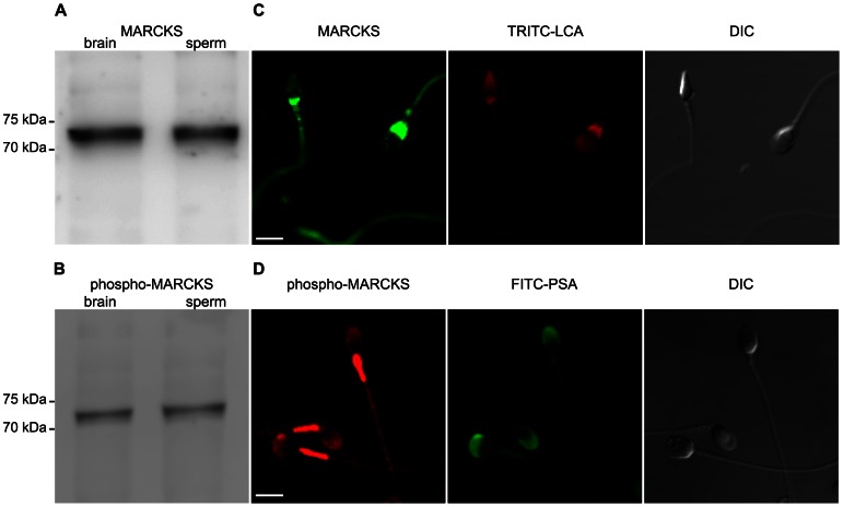
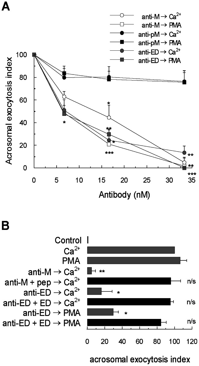
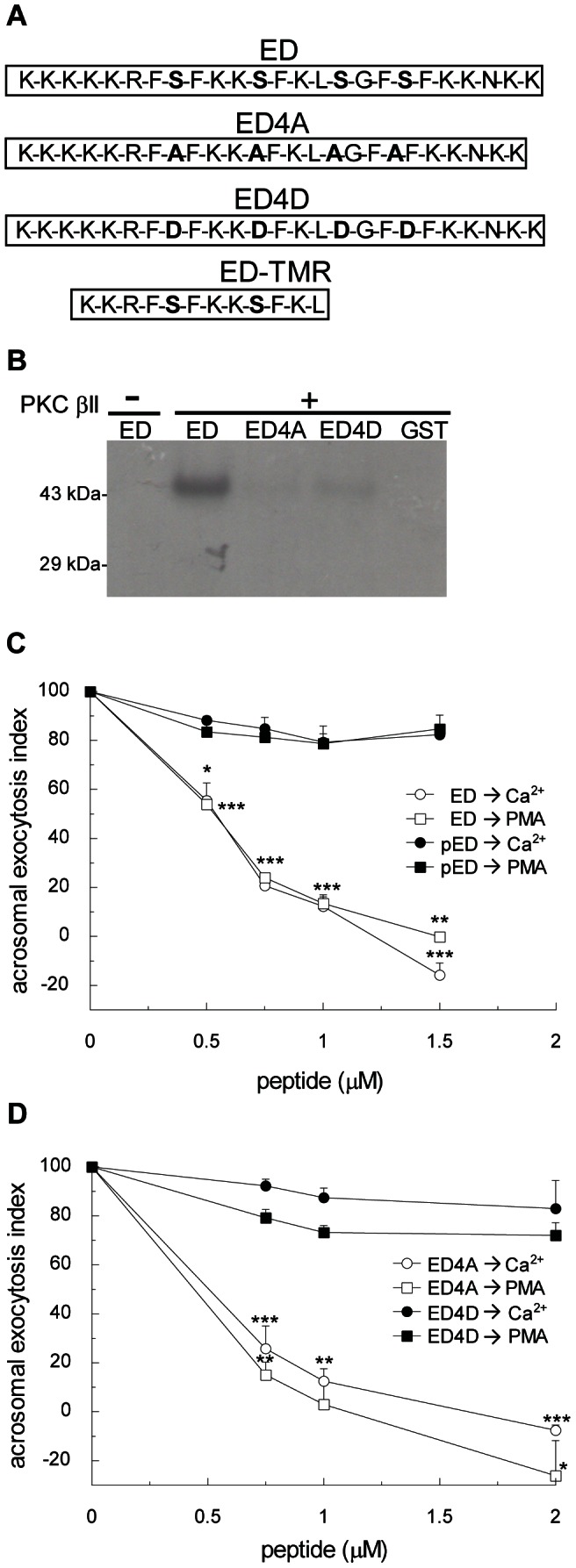
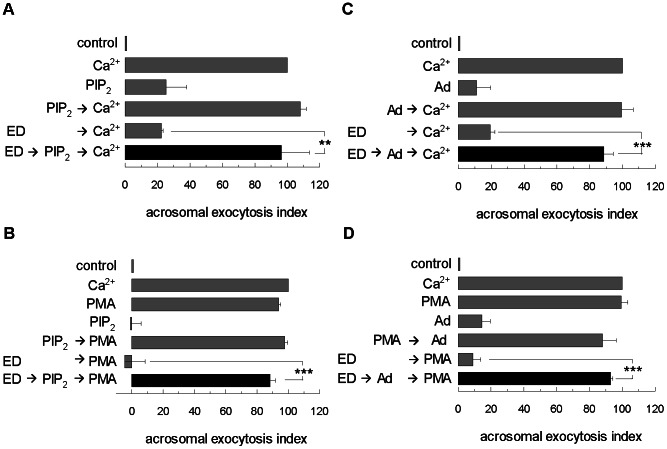
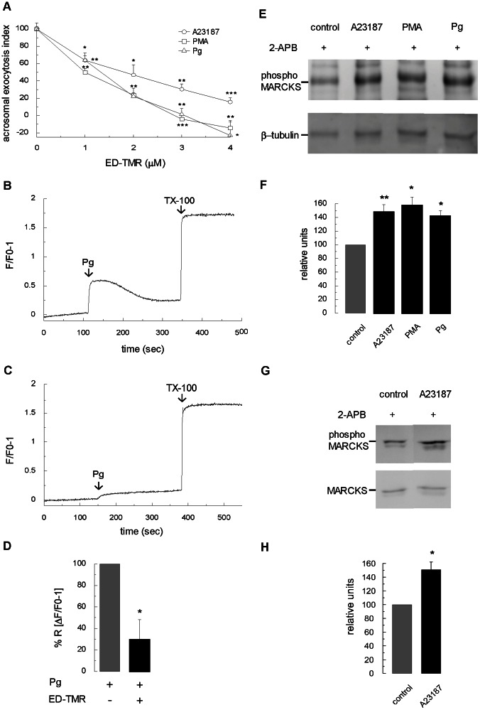
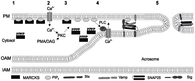
Acrosomal exocytosis is a calcium-regulated exocytosis that can be triggered by PKC activators. The involvement of PKC in acrosomal exocytosis has not been fully elucidated, and it is unknown if MARCKS, the major substrate for PKC, participates in this exocytosis. Here, we report that MARCKS is expressed in human spermatozoa and localizes to the sperm head and the tail. Calcium- and phorbol ester-triggered acrosomal exocytosis in permeabilized sperm was abrogated by different anti-MARCKS antibodies raised against two different domains, indicating that the protein participates in acrosomal exocytosis. Interestingly, an anti-phosphorylated MARCKS antibody was not able to inhibit secretion. Similar results were obtained using recombinant proteins and phospho-mutants of MARCKS effector domain (ED), indicating that phosphorylation regulates MARCKS function in acrosomal exocytosis. It is known that unphosphorylated MARCKS sequesters PIP2. This phospholipid is the precursor for IP3, which in turn triggers release of calcium from the acrosome during acrosomal exocytosis. We found that PIP2 and adenophostin, a potent IP3-receptor agonist, rescued MARCKS inhibition in permeabilized sperm, suggesting that MARCKS inhibits acrosomal exocytosis by sequestering PIP2 and, indirectly, MARCKS regulates the intracellular calcium mobilization. In non-permeabilized sperm, a permeable peptide of MARCKS ED also inhibited acrosomal exocytosis when stimulated by a natural agonist such as progesterone, and pharmacological inducers such as calcium ionophore and phorbol ester. The preincubation of human sperm with the permeable MARCKS ED abolished the increase in calcium levels caused by progesterone, demonstrating that MARCKS regulates calcium mobilization. In addition, the phosphorylation of MARCKS increased during acrosomal exocytosis stimulated by the same activators. Altogether, these results show that MARCKS is a negative modulator of the acrosomal exocytosis, probably by sequestering PIP2, and that it is phosphorylated during acrosomal exocytosis.


Conflict of interest statement

Competing Interests: The authors have declared that no competing interests exist.

Figures

Figure 1
Figure 1. MARCKS is expressed in human sperm.
(A) Proteins from whole sperm homogenates (sperm, 5×10⁶ cells) were resolved in 10% SDS-PAGE gels and immunoblotted with the anti-MARCKS antibody raised against the N-terminus of MARCKS. Brain extracts (brain) was used as a positive control. (B) Identical to A but immunoblotted with the anti-phospho-MARCKS antibody. (C) Sperm were double-stained with the anti-MARCKS antibody followed by an anti-mouse-DyLight488-conjugated antibody and with tetramethylrhodamine isothiocyanate-labeled Lens culinaris agglutinin to stain acrosomes (TRITC-LCA). (D) Sperm were double-stained with the anti-phospho-MARCKS antibody followed by an anti-rabbit-Cy3 antibody and FITC-coupled Pisum sativum agglutinin to stain acrosomes (FITC-PSA). Shown are representative images from three different experiments. DIC, cells observed with differential interference contrast. Bars = 5 µm
Figure 2
Figure 2. MARCKS participates in acrosomal exocytosis.
(A) Permeabilized sperm were incubated for 15 minutes at 37°C in the presence of increasing concentrations of anti-MARCKS N-19 antibody (anti-M, white symbols), anti-MARCKS ED (anti-ED, gray symbols) and anti-phospho-MARCKS (anti-pM, black symbols). Acrosomal exocytosis was initiated by adding 10 µM free Ca2+ (circles) or 200 nM PMA (squares). For all conditions, the incubation continued for an additional 15 minutes and acrosomes were evaluated by lectin binding. (B) Permeabilized sperm were incubated for 15 minutes at 37°C in the presence of 33 nM anti-MARCKS N-19 antibody (anti-M) preincubated with an excess (1∶ 10) of blocking peptide MARCKS N-19 (anti-M+pep) and in the presence of 33 nM anti-MARCKS ED antibody (anti-ED) preincubated with an excess (1∶10) of MARCKS effector domain (anti-ED+ED). Acrosomal exocytosis was then initiated by adding 10 µM free Ca2+ or 200 nM PMA and the incubation continued for an additional 15 minutes (black bar). Control experimental conditions (gray bars) include background acrosomal exocytosis in the absence of any stimulation (control), acrosomal exocytosis stimulated by 10 µM free Ca2+ (Ca2+) and by 200 nM PMA (PMA) and the effect of antibodies without peptide preincubation anti-MARCKS N-19 (anti-M) and anti-MARCKS ED (anti-ED). The percentage of reacted sperm was normalized as described in Materials and Methods. The data represent the means±SEM of at least three independent experiments. The asterisks indicate significant differences from similar conditions stimulated with Ca2+ (**, p<0.01; *, p< 0.05); n/s, not significant difference.
Figure 3
Figure 3. MARCKS inhibits acrosomal exocytosis and this effect is dependent on phosphorylation.
(A) Amino acid sequence (residues 145–169) of mouse wild type MARCKS effector domain (ED), the non-phosphorylable MARCKS ED mutant (ED4A), the phosphomimetic MARCKS ED mutant (ED4D), and permeable tetramethylrhodamine-conjugated MARCKS ED peptide (ED-TMR). (B) Purified GST fusion proteins of wild type MARCKS ED (ED), MARCKS ED4A mutant (ED4A), MARCKS ED4D (ED4D), and GST (26 kDa) were incubated for 40 minutes at 37°C with (+) or without (-) PKC βII under activating conditions in the presence of [γ32P]ATP. Samples were then resolved by 10% SDS-PAGE gels, and radiolabeled proteins were detected by autoradiography. Shown is a representative gel from three independent experiments. (C) Permeabilized sperm were treated for 15 minutes at 37°C with increasing concentrations of MARCKS effector domain (ED, white symbols) or in vitro phosphorylated MARCKS ED (pED, black symbols). Acrosomal exocytosis was initiated by adding 10 µM free Ca2+ (circles) or 200 nM PMA (squares). In all conditions, the incubation continued for an additional 15 minutes and acrosomal exocytosis was evaluated by lectin binding. (D) Permeabilized sperm were treated for 15 minutes at 37°C with increasing concentrations of MARCKS ED4A (ED4A, white symbols) or MARCKS ED4D (ED4D, black symbols). Acrosomal exocytosis was initiated by adding 10 µM free Ca2+ (circles) or 200 nM PMA (squares). In all conditions the incubation continued for an additional 15 minutes and acrosomal exocytosis was evaluated by lectin binding. In C and D, the percentage of reacted sperm was normalized as described in Materials and Methods. The data represent the means±SEM of at least three independent experiments. The asterisks indicate significant differences from similar conditions stimulated with Ca2+ (*, p<0.05; **, p<0.01; ***, p<0.001).
Figure 4
Figure 4. PIP2 and adenophostin rescue inhibition of MARCKS effector domain in acrosomal exocytosis.
Permeabilized sperm were treated for 15 minutes at 37°C with 1 µM MARCKS effector domain (ED); then, 50 µM PIP2 was added for another 15 min and acrosomal exocytosis was initiated by adding 10 µM free Ca2+ (A) or 200 nM PMA (B). The incubation continued for an additional 15 minutes (black bars). Control experimental conditions (gray bars) include background acrosomal exocytosis in the absence of any stimulation (control), acrosomal exocytosis stimulated by 10 µM free Ca2+ (Ca2+) and by 200 nM PMA (PMA), the absence of effect of PIP2 with or without acrosomal exocytosis stimulation. Permeabilized sperm were treated for 15 minutes at 37°C with 1 µM MARCKS effector domain (ED); then, 5 µM adenophostin was added (Ad), and acrosomal exocytosis was initiated by adding 10 µM free Ca2+ (C) or 200 nM PMA (D). The incubation continued for an additional 15 minutes (black bars). Control experimental conditions (gray bars) include background acrosomal exocytosis in the absence of any stimulation (control), acrosomal exocytosis stimulated by 10 µM free Ca2+ (Ca2+) and by 200 nM PMA (PMA), the absence of effect of adenophostin with or without acrosomal exocytosis stimulation, and the inhibitory effect of ED in acrosomal exocytosis. The percentage of reacted sperm was normalized as described in Materials and Methods. The data represent the means±SEM of at least three independent experiments. The asterisks indicate significant differences from similar conditions without PIP2 or adenophostin (**, p<0.01; ***, p<0.001).
Figure 5
Figure 5. Assessing MARCKS function on acrosomal exocytosis in non-permeabilized sperm.
(A) Capacitated and non-permeabilized sperm were treated for 30 minutes at 37°C with increasing concentrations of a permeable tetramethylrhodamine-labeled MARCKS ED peptide (ED-TMR). Acrosomal exocytosis was then initiated by adding 10 µM calcium ionophore A23187 (circles), 200 nM PMA (squares) or 15 µM progesterone (triangles), and the incubation continued for 15 minutes. The percentage of reacted sperm was normalized as described in Materials and Methods. The data represent the means±SEM of at least three independent experiments. The asterisks indicate significant differences from similar conditions without ED-TMR (*, p<0.05; **, p<0.01; ***, p<0.001). (B) Capacitated and non-permeabilized sperm were loaded with 2 µM Fluo-3-AM and incubated for 30 min at 37°C. At the indicated time (arrow) 15 µM progesterone was added. Maximal [Ca2+]i response was calibrated with 0.1% Triton X-100 (TX-100, arrow) at the end of the incubation period. Shown are traces representative of three experiments. The increase in fluorescence is expressed as (F/F0)-1 ((maximum fluorescence intensity/initial fluorescence)-1) versus time in seconds. (C) Identical to B with previous incubation for 30 min with 4 µM ED-TMR, before adding progesterone (arrow). (D) Quantification of three independent experiments. Bars represent mean±SEM; data were normalized to the calcium response to progesterone in the absence of ED-TMR. The asterisk indicates a significant difference from stimulation with progesterone (*, p<0.05; Student's t test). (E) Non-permeabilized and capacitated sperm were incubated for 15 min at 37°C with 100 µM 2-APB (control) and then treated for 20 min with 10 µM A23183, 200 nM PMA, or 15 µM progesterone. After treatment, proteins of each condition (5×10⁶ cells) were resolved in a 10% gels and transferred to PVDF membranes. Phosphorylated MARCKS was detected by Western blot assay using an anti-phospho-MARCKS antibody. Anti-β-tubulin antibody was used as loading control. (F) Quantification of three independent experiments showed in E. Bars represent mean±SEM; data were normalized against β-tubulin signal in control sperm. The asterisks indicate a significant difference from control sperm (*, p<0.05; **, p<0.01). (G) Capacitated and non-permeabilized sperm were incubated for 15 min at 37°C with 100 µM 2-APB (control) and then treated for 20 min with 10 µM A23183. After treatment, proteins of each condition (5×10⁶ cells) were resolved in a 10% gels and transferred to PVDF membranes. Phosphorylated MARCKS was detected by Western blot assay using an anti-phospho-MARCKS antibody. Anti-MARCKS antibody was used as loading control. (H) Quantification of three independent experiments showed in G. Bars represent mean±SEM; data were normalized against MARCKS signal in control sperm. Asterisk indicates a significant difference from control sperm (*, p<0.05).
Figure 6
Figure 6. Working model for the role of MARCKS in acrosomal exocytosis.
In resting sperm, MARCKS is present in two forms, a phosphorylated form in cytosol and another non-phosphorylated form, which binds to membranes sequestering PIP2 (1). When sperm is activated by a natural inducer such as progesterone, acrosomal exocytosis is triggered by a cytoplasmic increase of Ca2+ (2). Then, Ca2+ and DAG activate conventional PKC. Activated PKC phosphorylates the effector domain of MARCKS. Phosphorylated MARCKS is translocated to cytosol, augmenting the availability of PIP2 (3). PIP2 is hydrolyzed by PLC to generate IP3 and DAG. IP3 elicits the efflux of Ca2+ from acrosome (4). In addition, PIP2 interacts with SNAREs proteins and synaptotagmin (Stg) to fuse plasma and outer acrosomal membranes (5). Stx, syntaxin; VAMP, vesicle associated membrane protein; SNAP-25, synaptosomal-associated protein 25.

Similar articles

Cited by

References

    1. Yanagimachi R (1994) Mammalian fertilization. In: Knobil, E., Neill, J.D. (Eds.), The Physiology of Reproduction. Raven Press. In. pp. 189–281.
    1. Breitbart H (2002) Intracellular calcium regulation in sperm capacitation and acrosomal reaction. Mol Cell Endocrinol 187: 139–144. - PubMed
    1. Sistina Y, Lin M, Mate KE, Rodger JC (1993) Induction of the marsupial sperm acrosome reaction in vitro by treatment with diacylglycerols. J Reprod Fertil 99: 335–341. - PubMed
    1. De Jonge CJ, Han HL, Mack SR, Zaneveld LJ (1991) Effect of phorbol diesters, synthetic diacylglycerols, and a protein kinase C inhibitor on the human sperm acrosome reaction. J Androl 12: 62–70. - PubMed
    1. Roldan ER, Fragio C (1994) Diradylglycerols stimulate phospholipase A2 and subsequent exocytosis in ram spermatozoa. Evidence that the effect is not mediated via protein kinase C. Biochem J 297 (Pt 1): 225–232. - PMC - PubMed

Publication types

MeSH terms

Substances

LinkOut - more resources