. 2013 May 21;8(5):e64769.
doi: 10.1371/journal.pone.0064769. Print 2013.

Mechanical compliance and immunological compatibility of fixative-free decellularized/cryopreserved human pericardium

Affiliations

Mechanical compliance and immunological compatibility of fixative-free decellularized/cryopreserved human pericardium

Maria Cristina Vinci et al. PLoS One. .

Abstract

Background: The pericardial tissue is commonly used to produce bio-prosthetic cardiac valves and patches in cardiac surgery. The procedures adopted to prepare this tissue consist in treatment with aldehydes, which do not prevent post-graft tissue calcification due to incomplete xeno-antigens removal. The adoption of fixative-free decellularization protocols has been therefore suggested to overcome this limitation. Although promising, the decellularized pericardium has not yet used in clinics, due to the absence of proofs indicating that the decellularization and cryopreservation procedures can effectively preserve the mechanical properties and the immunologic compatibility of the tissue.

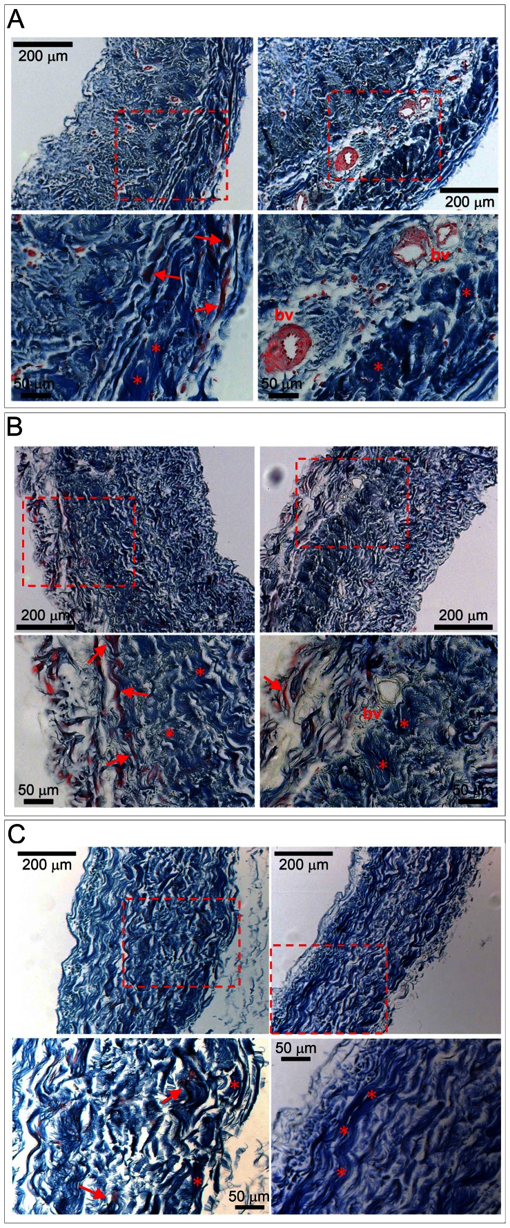
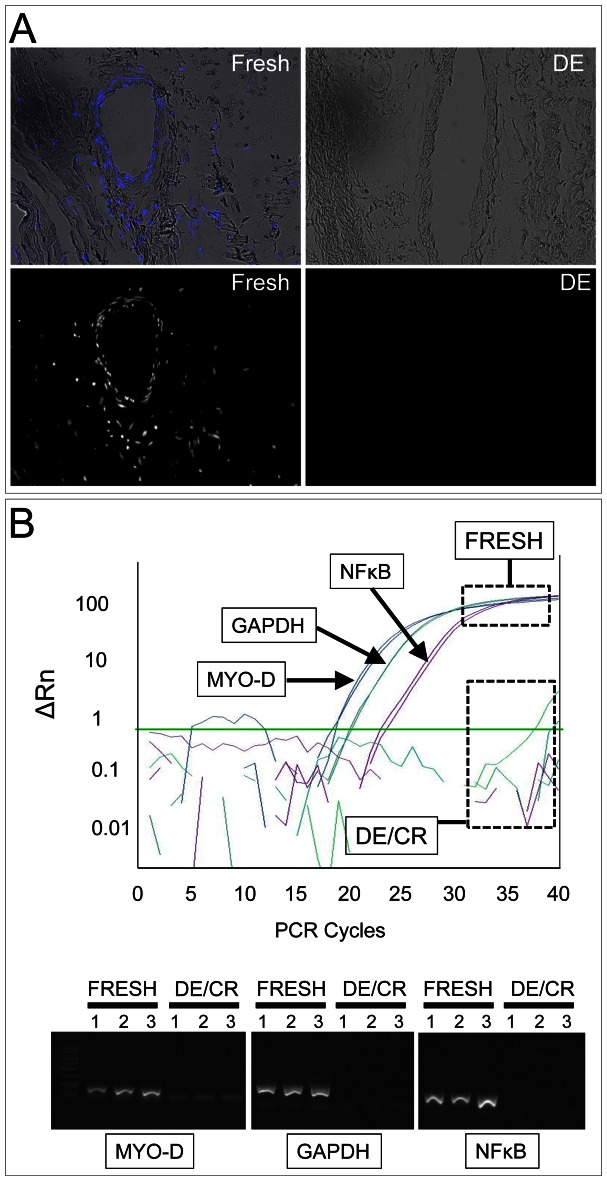
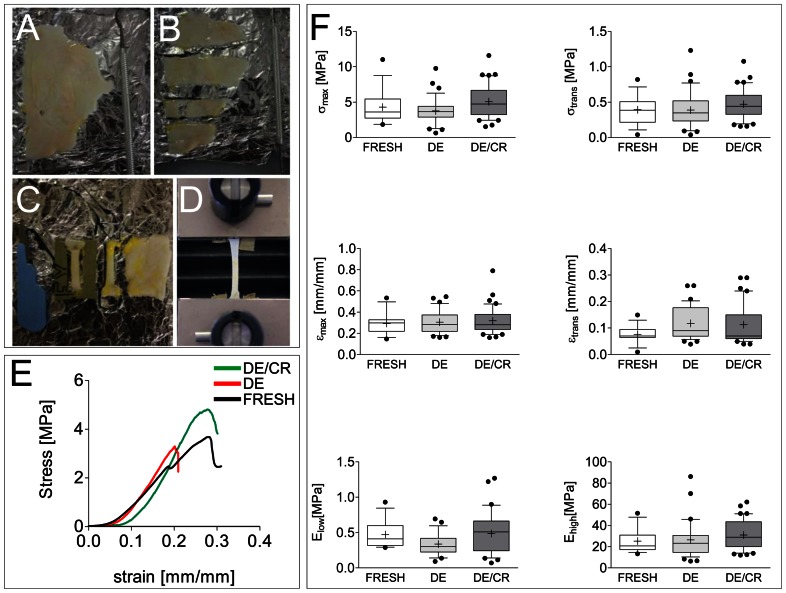
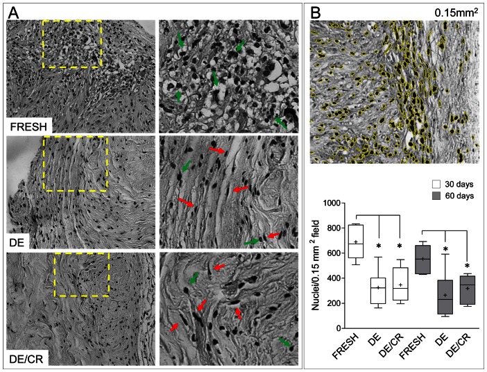
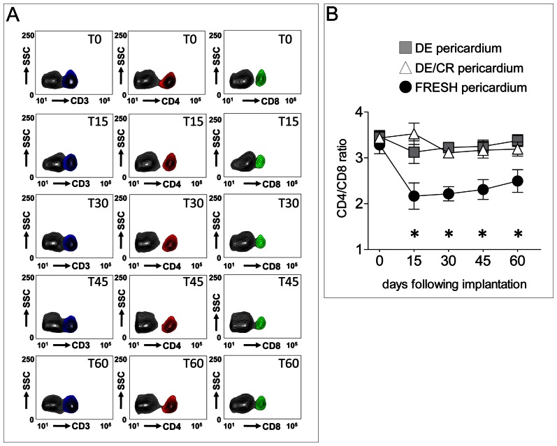
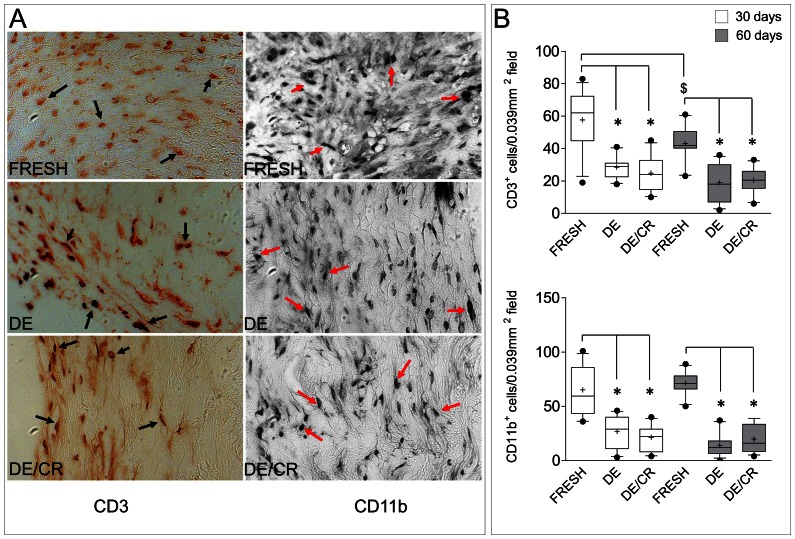
Principal findings: The aim of the present work was to validate a procedure to prepare decellularized/cryopreserved human pericardium which may be implemented into cardiovascular homograft tissue Banks. The method employed to decellularize the tissue completely removed the cells without affecting ECM structure; furthermore, uniaxial tensile loading tests revealed an equivalent resistance of the decellularized tissue to strain, before and after the cryopreservation, in comparison with the fresh tissue. Finally, immunological compatibility, showed a minimized host immune cells invasion and low levels of systemic inflammation, as assessed by tissue transplantation into immune-competent mice.

Conclusions: Our results indicate, for the first time, that fixative-free decellularized pericardium from cadaveric tissue donors can be banked according to Tissue Repository-approved procedures without compromising its mechanical properties and immunological tolerance. This tissue can be therefore treated as a safe homograft for cardiac surgery.


Conflict of interest statement

Competing Interests: Maurizio Pesce is a PLOS ONE Editorial Board member. This does not alter the authors' adherence to all the PLOS ONE policies on sharing data and materials.

Figures

Figure 1
Figure 1. Histological appearance and decellularization of pericardial samples.
(A, B, C) Masson's trichrome staining of the pericardial tissue before (A), after decellularization (B) and after decellularization/cryopreservation (C). Upper pictures in each panel show low magnification of the tissue, while lower pictures show magnification of the boxed areas. In these pictures it is evident that decellularization did not affect the structure of collagen bundles (*) and of elastic fibers (arrows). Bv: blood vessels.
Figure 2
Figure 2. Efficiency of the decellularization process.
(A) Hoechst 33258 staining was used to histologically assess the performance of the decellularization process. The upper panels show the overlapped bright field and Hoechst dye channels of the fresh and DE tissues without counterstaining, while lower panels show the dark field image of the Hoechst dye channel only in the same microscopic field. The lack of any signal in DE samples indicates a complete removal of double strand DNA from the tissue. (B) Q-PCR on DNA extracted from fresh and DE/CR samples shows complete removal of DNA by decellularization procedure using primers specific for the hMYO-D, hGAPDH and hNFκB genes promoter/coding sequences. The upper panel shows a representative amplification plot from one of the three FRESH samples with each primers pair, while the curves under the green bar are under-threshold signals generated by the corresponding DE/CR sample. The lower panel shows an agarose gel run of the PCR amplification products; as expected, no amplification bands were obtained by none of the DE/CR samples.
Figure 3
Figure 3. Uniaxial mechanical testing of fresh, DE and DE/CR pericardial samples.
(A–D) Preparation steps of pericardial specimens; intact pericardium (A), strips of pericardium (B), dog-shaped specimens obtained from the strips (C), specimen mounted into the clamps of the uniaxial test machine (D). (E) Representative curves showing the stress/strain relationship of fresh, DE and DE/CR samples. (F) Graphical representation of the six mechanical parameters obtained for fresh, DE and DE/CR samples: elastic modulus at low (Elow) and high (Ehigh) strain values, transition stress (σtrans) and strain (εtrans), maximum tensile stress (σmax) and strain (εmax). Results are graphically represented with inter-quartile range box plots and whiskers indicating 10th and 90th percentile, plus outlier values (•). Continuous lines in box plots indicate median values of each data set, while crosses indicate mean of the same data. None of these values in DE or DE/CR samples were significantly different from fresh tissue values as verified by Kruskall-Wallis test (n = 15, fresh samples; n = 36, DE samples; n = 41, DE/CR samples; pericardial tissues from 3 independent cadaveric donors).
Figure 4
Figure 4. Gross morphology and host cell invasion quantitative analysis at 30 and 60 days following fresh, DE and DE/CR tissues implantation into imunocompetent mice.
(A) Left panels show low magnification of fresh, DE and DE/CR pericardium after recovery from the subcutaneous position at 60 days. It is evident an overall higher number of cells in fresh vs. DE and DE/CR samples. Foci of inflammation with signs of tissue absorption were often observed in fresh samples, while these were never found in both DE and DE/CR tissues, indicating lower rejection. Panels on the right show high magnification of the yellow-boxed areas in left panels. It is evident that in fresh samples, infiltration was mainly due to cells with round nuclei, resembling inflammatory cells (green arrows), while the majority of infiltrating cells in DE and DE/CR pericardium specimens had elongated nuclei, resembling fibroblasts (red arrows). (B) Quantification of infiltrating cells was performed by computer assisted nuclei counting after appropriate filtering of the B/W images. Picture at the top is a 0.15 mm2 micrograph where nuclei were automatically recognized and contoured in yellow by Image-J software; box-plot in the bottom indicates the results of nuclei counting at 30 and 60 days in fresh, DE and DE/CR samples. Results are graphically represented with inter-quartile range box plots and whiskers indicating 10th and 90th percentile. Continuous lines in box plots indicate median values of each data set, while crosses indicate mean of the same data. * indicates P<0.05 for statistical comparison of fresh vs. DE and DE/CR samples at both time points by Kruskall-Wallis test with Dunn's post-hoc analysis (5≤n≤8).
Figure 5
Figure 5. Dynamics of CD3+, CD4+ and CD8+ cells mobilization in mice implanted with fresh, DE and DE/CR samples.
(A) Representative flow cytometry detection of CD3+ (left contour plots column), CD4+ (center contour plots column) and CD8+ (right contour plots column) circulating cells in a mouse receiving a non decellularized (FRESH) pericardial sample at 0, 15, 30, 45 and 60 days. CD3+, CD4+ and CD8+ cells are indicated, respectively by blue, red and green contours on the right of the main PBMNCs population (grey contours), established by staining with isotype control antibodies. (B) CD4+/CD8+ lymphocytes ratio was determined at all the time points and plotted. Mice receiving fresh pericardium showed a reduced ratio suggesting increase in the number of circulating T-lymphocytes implicated in T cell-mediated tissue rejection. * indicate a statistical difference (P<0.05, by two-ways ANOVA with Bonferroni post-hoc test, n = 5) between the CD4/CD8 ratio in mice receiving fresh vs. those implanted with DE and DE/CR tissues.
Figure 6
Figure 6. Infiltration of the implanted pericardial tissue by CD3+ lymphocytes and CD11b+ macrophages.
(A) Representative images of fresh, DE and DE/CR pericardium tissue sections recovered by a mouse at 60 days and stained with CD3− and CD11b-specific antibodies. Black arrows and red arrows indicate, respectively CD3+ and CD11b+ cells present in the tissue. Note the higher number of cells in fresh pericardial specimens compared to DE or DE/CR samples. (B) Quantification of CD3+ (upper plot) and CD11b+ cells (lower plot) by software-assisted manual counting (Image J) into fixed (0.039 mm2) tissue sections areas. At both time points the number of CD3+ and CD11b+ cells was significantly higher in mice receiving fresh vs. DE or DE/CR pericardium (* indicate P<0.05 by 1-way ANOVA with Newman-Keuls post-hoc; n = 12). Interestingly, the number of lymphocytes was significantly lower at 60 days compared with 30 days in mice receiving fresh pericardium ($ indicates P<0.05 by Mann-Whitney rank sum test; n = 12).

References

    1. Carpentier A, Lemaigre G, Robert L, Carpentier S, Dubost C (1969) Biological factors affecting long-term results of valvular heterografts. J Thorac Cardiovasc Surg 58: 467–483. - PubMed
    1. Grabenwoger M, Sider J, Fitzal F, Zelenka C, Windberger U, et al. (1996) Impact of glutaraldehyde on calcification of pericardial bio-prosthetic heart valve material. Ann Thorac Surg 62: 772–777. - PubMed
    1. Vincentelli A, Latremouille C, Zegdi R, Shen M, Lajos PS, et al. (1998) Does glutaraldehyde induce calcification of bio-prosthetic tissues? Ann Thorac Surg 66: S255–258. - PubMed
    1. Bloch O, Golde P, Dohmen PM, Posner S, Konertz W, et al. (2011) Immune response in patients receiving a bio-prosthetic heart valve: lack of response with decellularized valves. Tissue Eng Part A 17: 2399–2405. - PubMed
    1. Galili U (2005) The [alpha]-gal epitope and the anti-Gal antibody in xenotransplantation and in cancer immunotherapy. Immunol Cell Biol 83: 674–686. - PubMed

Publication types