. 2013 May 25:6:27.
doi: 10.1186/1756-6606-6-27.

Neurological characterization of mice deficient in GSK3α highlight pleiotropic physiological functions in cognition and pathological activity as Tau kinase

Affiliations

Neurological characterization of mice deficient in GSK3α highlight pleiotropic physiological functions in cognition and pathological activity as Tau kinase

Hervé Maurin et al. Mol Brain. .

Abstract

Background: GSK3β is involved in a wide range of physiological functions, and is presumed to act in the pathogenesis of neurological diseases, from bipolar disorder to Alzheimer's disease (AD). In contrast, the GSK3α isozyme remained largely ignored with respect to both aspects.

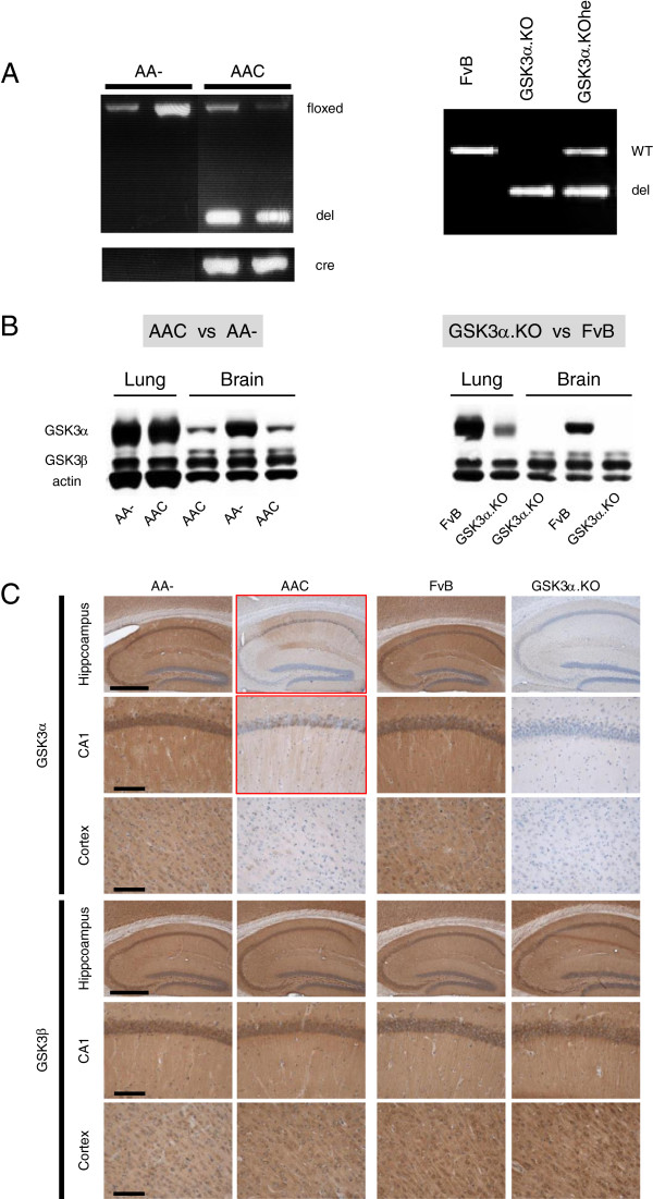
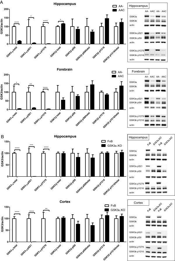
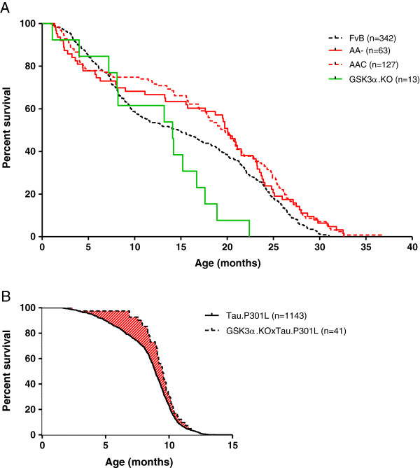
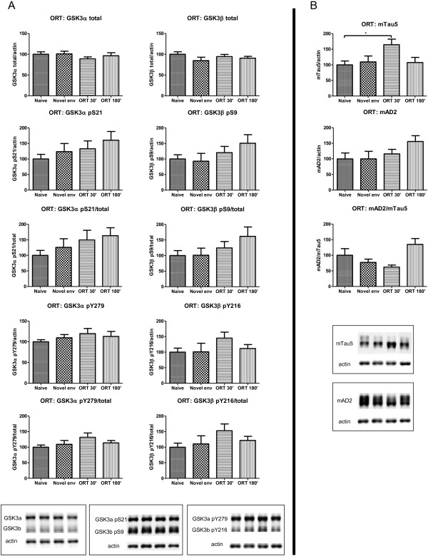
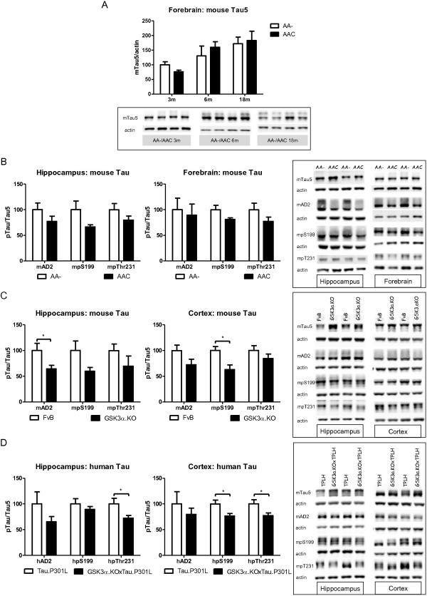
Results: We generated and characterized two mouse strains with neuron-specific or with total GSK3α deficiency. Behavioral and electrophysiological analysis demonstrated the physiological importance of neuronal GSK3α, with GSK3β not compensating for impaired cognition and reduced LTP. Interestingly, the passive inhibitory avoidance task proved to modulate the phosphorylation status of both GSK3 isozymes in wild-type mice, further implying both to function in cognition. Moreover, GSK3α contributed to the neuronal architecture of the hippocampal CA1 sub-region that is most vulnerable in AD. Consequently, practically all parameters and characteristics indicated that both GSK3 isoforms were regulated independently, but that they acted on the same physiological functions in learning and memory, in mobility and in behavior.

Conclusions: GSK3α proved to be regulated independently from GSK3β, and to exert non-redundant physiological neurological functions in general behavior and in cognition. Moreover, GSK3α contributes to the pathological phosphorylation of protein Tau.


Figures

Figure 1
Figure 1
Genotyping and characterization of neuron-specific and total GSK3α deficient mice. A. Genotyping by PCR reveals floxed or recombined (del) GSK3α alleles in AAC and AA- mice by the action of Cre-recombinase (Cre) (left panel). In GSK3α.KO mice (right panel) the same PCR reaction defines wild-type (WT) and recombined GSK3α alleles (del) in homozygous and heterozygous GSK3α.KO mice. B. Representative western blots for the GSK3 isozymes in total protein extracts of brain and lung from AA-, AAC, FvB and GSK3α.KO mice, as indicated. Note some residual GSK3α protein in the brain of AAC mice as opposed to the total absence in the GSK3α.KO mice. The minor non-specific reaction observed in extracts of lungs of GSK3α.KO mice was caused by a non-identified protein. C. Representative IHC of hippocampus, CA1 and cortex for either GSK3 isozyme on brain sections from both deficient genotypes and their respective control mice: AAC versus AA- mice and GSK3α.KO versus wild-type FvB mice. Note some residual GSK3α immunoreaction in the hippocampus of AAC mice, as opposed to total absence in GSK3α.KO mice (cfr text for details).
Figure 2
Figure 2
Biochemical analysis of GSK3 isozymes in mouse brain extracts. Levels of total GSK3 protein, pS21/S9 and pY279/Y216 in total protein extracts from hippocampus and forebrain from AAC mice compared to control AA- mice (A) and from hippocampus and cortex from GSK3α.KO mice versus wild-type FvB mice (B). Western blots were digitally quantified, normalized for actin and reported relative to the respective control mice. Data (mean±SEM) are statistically analyzed by unpaired Student’s t-test (two-tailed), n=6 or 7 per genotype; *p<0.05, ** p<0.01, *** p<0.001, **** p<0.0001.
Figure 3
Figure 3
Kaplan-Meier survival curves. Spontaneous death of AAC, AA-, GSK3α.KO and wild-type FvB mice (panel A) and in GSK3α.KOxTau.P301L bigenic mice versus the parental Tau.P301L mice (panel B) housed in the same conditions in our breeding colony. The number of mice of either gender is indicated. Note that earlier death of GSK3α.KO mice (panel A) is based on the relatively low number of mice that died spontaneously over the period of observation. The shaded area in panel B emphasizes the delayed mortality of young GSK3α.KOxTau.P301L mice, relative to the parental Tau.P301L mice.
Figure 4
Figure 4
Body weight and brain weight. Body weight, wet weight of heart, brain, hippocampus of (A) AAC and AA- mice of either gender at young (n=4/5) and older age (n=6/9) and (B) of GSK3α.KO, GSK3α.KOxTau.P301L, Tau.P301L and FvB mice (n=4/10) at young and older age. Data (mean±SEM) are statistically analyzed by unpaired Student’s t-test (two-tailed), * p<0.01, ** p<0.05, *** p<0.001, **** p<0.0001.
Figure 5
Figure 5
Male GSK3α.KO mice are infertile. A. Breeding performance of GSK3α.KO mice versus FvB mice, estimated as number of pups and number of litters over a period of 111 days from 5 breeding couples/genotype). B. Testis weight, sperm cell counting and motility index assessed in males (n=5). All data (mean±SEM) are statistically analyzed by unpaired Student’s t-test (two-tailed): Testis weight, * p=0.030; sperm cells, p=0.198; motility index, * p=0.032. The red symbols in panels A and B represent a sterile FvB male. C. Sperm cells from GSKα.KO and FvB wild-type mice. Inset: single sperm at higher magnification.
Figure 6
Figure 6
Reduced CA1 in neuron-specific and in total GSK3α-deficient mice. Left panels: ratio of CA1 pyramidal neurons to total CA1 area in (A) AA- and AAC mice (n=4; 3 sections/mouse) and (B) FvB and GSK3α.KO mice (n=8/9; 3 sections/mouse). Data (mean±SEM) are statistically analyzed by unpaired Student’s t-test (two-tailed), **** p<0.0001. Right panels: representative images of hippocampus and CA1 pyramidal layer (insets). Scale bars 400 μm; insets: 100 μm.
Figure 7
Figure 7
Electrophysiology of neuron-specific GSK3α deficient mice. A. Input–output curve generated by gradually increasing stimulus intensity in sections from AAC mice compared to control AA- mice (n=6). Two-way Anova; genotype: F(1,399)=15.20, *** p<0.0001; stimulation intensity: F(6,399)=106.9, p<0.0001; interaction: F(6,399)=1.256, p=0.2766. B. Paired pulse facilitation (PPF) induced in 4 intervals of 25, 50, 100 and 200 ms in CA1 stratum radiatum of sections from AAC mice compared to AA- mice (n=6). Data are expressed as the ratio of the second to the first fEPSP slope. Two-way Anova; genotype: F(1,220)=1.124, p=0.2903; time: F(3,220)=34.35, p<0.0001; interaction: F(3,220)=1.685, p=0.1710. C. Synaptic fatigue analyzed for 1 to 10 stimulations in sections from AAC mice compared to AA- mice (n=6). Two-way Anova; genotype: F(1,320)=67.23, **** p<0.0001; stimulus: F(9,320)=321.3, p<0.0001; Interaction: F(9,320)=1.030, p=0.4153. D. LTP was recorded in CA1 stratum radiatum in sections from AAC and AA- mice (n=6). L-LTP was analyzed during the last 2 hours of recording. Unpaired Student’s t-test (two-tailed); *p=0.0369.
Figure 8
Figure 8
Biochemical repercussions of PIA on GSK3 and protein tau in hippocampus of wild-type mice. A. Biochemical analysis of GSK3 by western blotting of total protein extracts from hippocampus of FvB wild-type mice (n=10) at different timepoints before or after they performed the PIA task (n=6 per timepoint). All data normalized to actin and reported relative to naive control mice. Data (mean±SEM) are statistically analyzed by one-way Anova (Dunnet’s post hoc test compared to naive). GSK3α: F(3,24)=0.5422; p=0.6581. GSK3β: F(3,24)=1.523; p=0.2339. GSK3α pS21: F(3,24)=12.58; p<0.0001. GSK3α pS21/total: F(3,24)=12.11; p<0.0001. GSK3β pS9: F(3,24)=5.240; p=0.0063. GSK3β pS9/total: F(3,24)=4.832; p=0.0090. GSK3α pY279: F(3,24)=4.973; p=0.0080. GSK3α pY279/total: F(3,24)=3.346; p=0.0359. GSK3β pY216: F(3,24)=3.875; p=0.0216. GSK3β pY216/total: F(3,24)=4.528; p=0.0119. * p<0.05; ** p<0.01; *** p<0.001. Lower panels show representative western blots. B. Biochemical analysis of protein Tau by western blotting of total protein extracts from hippocampus of FvB wild-type mice (n=10) at different timepoints before or after they performed the PIA task (n=6 per timepoint). All data normalized to actin and reported relative to naive control mice. Data (mean±SEM) are statistically analyzed by one-way Anova (Dunnet’s post hoc test compared to naive). Tau5: F(3,24)=7.625; p=0.0009. AD2: F(3,24)=5.162; p=0.0068. AD2/Tau5: F(3,24)=1.761; p=0.1815. * p<0.05; ** p<0.01; *** p<0.001. Lower panels show representative western blots.
Figure 9
Figure 9
Biochemical repercussions of NORT on GSK3 and Tau in hippocampus of wild-type mice. A. Biochemical analysis of GSK3 by western blotting of total protein extracts from hippocampus of FvB wild-type mice (n=10) at difference timepoints before or after they performed the NORT task (n=6 per timepoint). All data normalized to actin and reported relative to naive control mice. Data (mean±SEM) are statistically analyzed by one-way Anova (Dunnet’s post hoc test compared to naive). GSK3α: F(3,24)= 0.6609; p=0.5842. GSK3β: F(3,24)= 1.131; p=0.3566. GSK3α pS21: F(3,24)= 1.351; p=0.2816. GSK3α pS21/total: F(3,24)= 1.529; p=0.2327. GSK3β pS9: F(3,24)= 1.469; p=0.2479. GSK3β pS9/total: F(3,24)= 1.699; p=0.1939. GSK3α pY279: F(3,24)= 1.002; p=0.4091. GSK3α pY279/total: F(3,24)= 1.871; p=0.1614. GSK3β pY216: F(3,24)= 1.302; p=0.2967. GSK3β pY216/total: F(3,24)= 1.580; p=0.2203. Lower panels show representative western blots. B. Biochemical analysis of protein Tau by western blotting of total protein extracts from hippocampus of FvB wild-type mice (n=10) at difference timepoints before or after they performed the NORT task (n=6 per timepoint). All data normalized to actin and reported relative to naive control mice. Data (mean±SEM) are statistically analyzed by one-way Anova (Dunnet’s post hoc test compared to naive). Tau5: F(3,24)= 3.286; p=0.0380. AD2: F(3,24)= 1.637; p=0.2071. AD2/Tau5: F(3,24)= 2.593; p=0.0761. * p<0.05. Lower panels show representative western blots.
Figure 10
Figure 10
Biochemical analysis of protein Tau in GSK3α deficient mice. A. Biochemical analysis by western blotting of total protein extracts from forebrain of AAC and AA- mice aged 3, 6 and 18 months (n=5/age). Tau protein levels are normalized to actin and expressed relative to control AA- mice at age 3 months. Data (mean±SEM) are statistically analyzed by two-way Anova (Bonferroni post hoc test), genotype: F(1,24)=3.40, p=0.5093; age: F(2,24)=37.60, p=0.0027; interaction: F(2,24)=0.20, p=0.7759. Lower panels show representative western blots. B. Biochemical analysis by western blotting for phospho-epitopes pS396/404, pS199 and pT231 of endogenous mouse Tau in total protein extracts from hippocampus and forebrain of AAC and control AA- mice. C. Biochemical analysis by western blotting for phospho-epitopes pS396/404, pS199 and pT231 of endogenous mouse Tau in total protein extracts from hippocampus and cortex of GSK3α.KO and FVB wild-type mice. D. Biochemical analysis by western blotting for phospho-epitopes pS396/404, pS199 and pT231 of human Tau.P301L in total protein extracts from hippocampus and cortex of GSK3α.KOxTau.P301L mice and the parental Tau.P301L mice. In panels B-D, all data are normalized for total Tau and reported relative to the respective control mice. Data (mean±SEM) are statistically analyzed by unpaired Student’s t-test (two-tailed), n=6 or 7 per genotype; * p<0.05.

Similar articles

Cited by

References

    1. Patel S, Doble B, Woodgett JR. Glycogen synthase kinase-3 in insulin and Wnt signalling: a double-edged sword? Biochem Soc Trans. 2004;32:803–808. doi: 10.1042/BST0320803. - DOI - PMC - PubMed
    1. Doble BW, Woodgett JR. GSK-3: tricks of the trade for a multi-tasking kinase. J Cell Sci. 2003;116:1175–1186. doi: 10.1242/jcs.00384. - DOI - PMC - PubMed
    1. Jope RS, Johnson GVW. The glamour and gloom of glycogen synthase kinase-3. Trends Biochem Sci. 2004;29:95–102. doi: 10.1016/j.tibs.2003.12.004. - DOI - PubMed
    1. Hanger DP, Noble W. Functional implications of glycogen synthase kinase-3-mediated tau phosphorylation. Int J Alzheimer’s Disease. 2011;2011:352805. - PMC - PubMed
    1. Kremer A, Louis JV, Jaworski T, Van Leuven F. GSK3 and Alzheimer’s disease: facts and fiction…. Front Mol Neurosci. 2011;4:1–10. - PMC - PubMed

Publication types

MeSH terms

LinkOut - more resources