Engineering three-dimensional collagen-IKVAV matrix to mimic neural microenvironment
- PMID: 23705903
- PMCID: PMC3750678
- DOI: 10.1021/cn400075h
Engineering three-dimensional collagen-IKVAV matrix to mimic neural microenvironment
Abstract
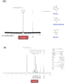
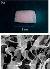
Engineering the cellular microenvironment has great potential to create a platform technology toward engineering of tissue and organs. This study aims to engineer a neural microenvironment through fabrication of three-dimensional (3D) engineered collagen matrixes mimicking in-vivo-like conditions. Collagen was chemically modified with a pentapeptide epitope consisting of isoleucine-lysine-valine-alanine-valine (IKVAV) to mimic laminin structure supports of the neural extracellular matrix (ECM). Three-dimensional collagen matrixes with and without IKVAV peptide modification were fabricated by freeze-drying technology and chemical cross-linking with glutaraldehyde. Structural information of 3D collagen matrixes indicated interconnected pores structure with an average pore size of 180 μm. Our results indicated that culture of dorsal root ganglion (DRG) cells in 3D collagen matrix was greatly influenced by 3D culture method and significantly enhanced with engineered collagen matrix conjugated with IKVAV peptide. It may be concluded that an appropriate 3D culture of neurons enables DRG to positively improve the cellular fate toward further acceleration in tissue regeneration.
Figures


Similar articles
-
Tailored laminin-332 alpha3 sequence is tethered through an enzymatic linker to a collagen scaffold to promote cellular adhesion.Acta Biomater. 2009 Sep;5(7):2441-50. doi: 10.1016/j.actbio.2009.03.018. Epub 2009 Mar 26. Acta Biomater. 2009. PMID: 19364681
-
Decellularized porcine brain matrix for cell culture and tissue engineering scaffolds.Tissue Eng Part A. 2011 Nov;17(21-22):2583-92. doi: 10.1089/ten.TEA.2010.0724. Epub 2011 Oct 17. Tissue Eng Part A. 2011. PMID: 21883047 Free PMC article.
-
Cell behavior on protein matrices containing laminin α1 peptide AG73.Biomaterials. 2011 Jul;32(19):4327-35. doi: 10.1016/j.biomaterials.2011.02.052. Epub 2011 Mar 21. Biomaterials. 2011. PMID: 21420730
-
Laminin-derived Ile-Lys-Val-ala-Val: a promising bioactive peptide in neural tissue engineering in traumatic brain injury.Cell Tissue Res. 2018 Feb;371(2):223-236. doi: 10.1007/s00441-017-2717-6. Epub 2017 Oct 30. Cell Tissue Res. 2018. PMID: 29082446 Review.
-
In vitro studies on the control of nerve fiber growth by the extracellular matrix of the nervous system.J Physiol (Paris). 1987;82(4):258-70. J Physiol (Paris). 1987. PMID: 3332689 Review.
Cited by
-
Collagen Film Activation with Nanoscale IKVAV-Capped Dendrimers for Selective Neural Cell Response.Nanomaterials (Basel). 2021 Apr 28;11(5):1157. doi: 10.3390/nano11051157. Nanomaterials (Basel). 2021. PMID: 33925197 Free PMC article.
-
Bioactivated Oxidized Polyvinyl Alcohol towards Next-Generation Nerve Conduits Development.Polymers (Basel). 2021 Sep 30;13(19):3372. doi: 10.3390/polym13193372. Polymers (Basel). 2021. PMID: 34641183 Free PMC article.
-
Research Progress of Chitosan-Based Biomimetic Materials.Mar Drugs. 2021 Jun 27;19(7):372. doi: 10.3390/md19070372. Mar Drugs. 2021. PMID: 34199126 Free PMC article. Review.
-
Hydrogels in Spinal Cord Injury Repair: A Review.Front Bioeng Biotechnol. 2022 Jun 21;10:931800. doi: 10.3389/fbioe.2022.931800. eCollection 2022. Front Bioeng Biotechnol. 2022. PMID: 35800332 Free PMC article. Review.
-
Nano-scaled MTCA-KKV: for targeting thrombus, releasing pharmacophores, inhibiting thrombosis and dissolving blood clots in vivo.Int J Nanomedicine. 2019 Jul 3;14:4817-4831. doi: 10.2147/IJN.S206294. eCollection 2019. Int J Nanomedicine. 2019. PMID: 31308660 Free PMC article.
References
-
- Baker S. C.; Atkin N.; Gunning P. A.; Granville N.; Wilson K.; Wilson D.; Southgatea J. (2006) Characterization of electrospun polystyrene scaffolds for three-dimensional in vitro biological studies. Biomaterials 27, 3136–3146. - PubMed
-
- Smith L. A.; Ma P. X. (2004) Nano-fibrous scaffolds for tissue engineering. Colloids Surf., B 10, 125–131. - PubMed
-
- Hosseinkhani H.; Hosseinkhani M.; Kobayashi H. (2006) Design of tissue engineered nanoscaffold through self assembly of peptide amphiphile. J. Bioact. Compat. Polym. 21, 277–296.
-
- Lennon D. P.; Haynesworth S. E.; Dennis J. E.; Caplan A. I. (1995) A chemically defined medium supports in vitro proliferation and maintains the osteochondral potential of rat marrow-derived mesenchymal stem cells. Exp. Cell Res. 219, 211–222. - PubMed
-
- Hosseinkhani H.; Hosseinkhani M.; Khademhosseini A. (2006) Tissue regeneration through self-assembled peptide amphiphile nanofibers. Yakhteh Med. J. 8, 204–209.
Publication types
MeSH terms
Substances
LinkOut - more resources
Full Text Sources
Other Literature Sources

