. 2013 May 25:14:25.
doi: 10.1186/1471-2121-14-25.

Syndecan-2 is upregulated in colorectal cancer cells through interactions with extracellular matrix produced by stromal fibroblasts

Affiliations

Syndecan-2 is upregulated in colorectal cancer cells through interactions with extracellular matrix produced by stromal fibroblasts

Carolina Meloni Vicente et al. BMC Cell Biol. .

Abstract

Background: The extracellular matrix (ECM) influences the structure, viability and functions of cells and tissues. Recent evidence indicates that tumor cells and stromal cells interact through direct cell-cell contact, the production of ECM components and the secretion of growth factors. Syndecans are a family of transmembrane heparan sulfate proteoglycans that are involved in cell adhesion, motility, proliferation and differentiation. Syndecan-2 has been found to be highly expressed in colorectal cancer cell lines and appears to be critical for cancer cell behavior. We have examined the effect of stromal fibroblast-produced ECM on the production of proteoglycans by colorectal cancer cell lines.

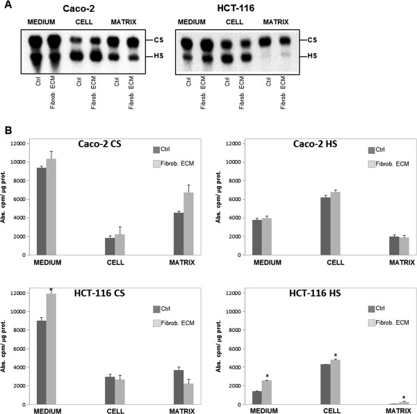
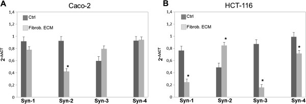
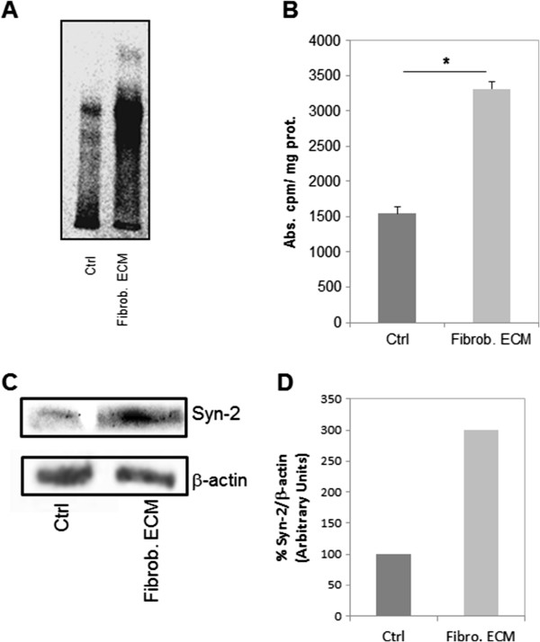
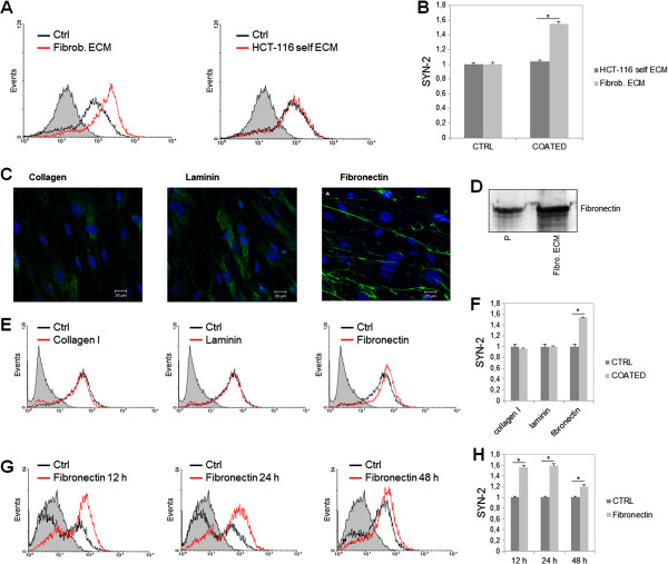
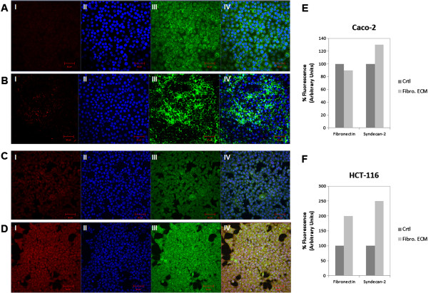
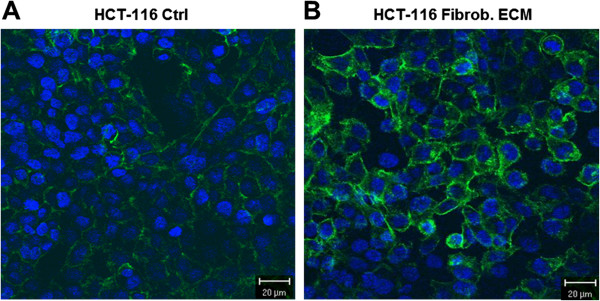
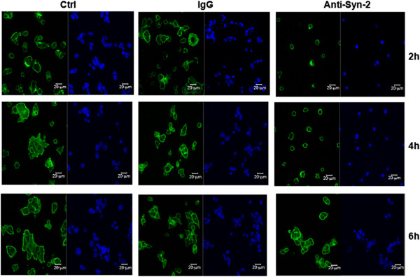
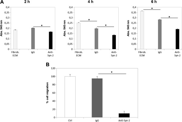
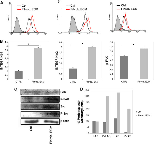
Results: Our results showed that in a highly metastatic colorectal cancer cell line, HCT-116, syndecan-2 expression is enhanced by fibroblast ECM, while the expression of other syndecans decreased. Of the various components of the stromal ECM, fibronectin was the most important in stimulating the increase in syndecan-2 expression. The co-localization of syndecan-2 and fibronectin suggests that these two molecules are involved in the adhesion of HCT-116 cells to the ECM. Additionally, we demonstrated an increase in the expression of integrins alpha-2 and beta-1, in addition to an increase in the expression of phospho-FAK in the presence of fibroblast ECM. Furthermore, blocking syndecan-2 with a specific antibody resulted in a decrease in cell adhesion, migration, and organization of actin filaments.

Conclusions: Overall, these results show that interactions between cancer cells and stromal ECM proteins induce significant changes in the behavior of cancer cells. In particular, a shift from the expression of anti-tumorigenic syndecans to the tumorigenic syndecan-2 may have implications in the migratory behavior of highly metastatic tumor cells.


Figures

Figure 1
Figure 1
Effect of stromal fibroblast ECM on the synthesis of GAGs by Caco-2 and HCT-116 cells. Cancer cells were cultured in the absence (Ctrl) or presence (Fibrob. ECM) of stromal fibroblast ECM. GAGs were labeled with [35S]Na2SO4 and were purified from the culture medium (MEDIUM), cancer cells (CELL) and the matrix (MATRIX) produced by Caco-2 or HCT-116 cells. (A) The content of GAGs from these compartments was analyzed by agarose gel electrophoresis in 1,3-diaminepropane acetate buffer (0.05-M pH 9.0). The gel was exposed to a screen and the bands were identified using an image analysis system, the Cyclone® Storage Phosphor System-Packard Instrument. (B) Quantification was performed by densitometry with Opti-Quanti Software. Heparan sulfate (HS), chondroitin sulfate (CS). *p ≤ 0.05 compared to control.
Figure 2
Figure 2
Effect of stromal fibroblast ECM on the expression of syndecans in Caco-2 and HCT-116 cells. Caco-2 (A) and HCT-116 cells (B) were cultured on Petri dishes in the absence (Ctrl) or presence of fibroblast ECM (Fibrob. ECM) for three days, and RNA was extracted. The expression level of each gene was normalized to that of β-actin. The data from each experiment were obtained in triplicate and are represented as the average ± standard deviation. *p ≤ 0.05. Syndecan-1 (Syn-1), syndecan-2 (Syn-2), syndecan-3 (Syn-3), syndecan-4 (Syn-4).
Figure 3
Figure 3
Expression of syndecan-2 in HCT-116 cells. (A) Immunoprecipitation of syndecan-2 from HCT-116 cells in the absence of matrix (Ctrl) or in the presence of stromal fibroblast ECM (Fibrob. ECM). HCT-116 cells were cultured for 72h and exposed to [35S]sulfate for 24 h, and the radioactive proteoglycans were extracted as described in Methods. The proteoglycans from the cells were immunoprecipitated with anti-syndecan-2 antibody and were then applied to the gel. (B) Quantification of the experiment shown in A. (C) HCT-116 cells were seeded on stromal fibroblast ECM and cultured for three days. Lysate proteins were separated on 10% SDS-PAGE and electro-transferred to PVDF membrane. Membranes were blocked and incubated using anti-syndecan-2 (Syn-2) and anti-β-actin (loading control). Antibody binding was visualized by chemiluminescence and the relative levels of these proteins were determined by densitometric analysis (D). *p ≤ 0.05 compared to control.
Figure 4
Figure 4
Flow cytometric analysis of syndecan-2 surface expression on HCT-116 cells. (A) HCT-116 cells were cultured for 48 h in the absence of matrix (Ctrl), in the presence of stromal fibroblast ECM (Fibrob. ECM) or in the presence of their own matrix (HCT-116 self ECM) and then immunostained with anti-syndecan-2 antibody. (B) The relative staining was determined by densitometric analysis. (C) Fibroblasts were immunostained with anti-fibronectin antibody, anti-laminin antibody or anti-collagen antibody to confirm the presence of these proteins. The photo shows images that were obtained using a confocal microscope. HCT-116 cells were cultured for 72 h on fibronectin, laminin or collagen I and labeled with anti-syndecan-2 antibody. (D) Fibroblasts were cultured in Petri dishes until confluence and its ECM was extracted as described in Methods. Total protein from the ECM (Fibro. ECM) was then applied to polyacrylamide gel 7.5% with 10μg of standard fibronectin (P). After transfer, the nitrocellulose membrane was incubated with anti-fibronectin and revealed with DAB. Collagen and laminin were not detected on ECM produced by fibroblasts through Western blotting. (E) HCT-116 cells were cultured in the presence of collagen-I, laminin or fibronectin for 72 h and then stained with anti-syndecan-2 antibody and the relative levels of this protein were determined by densitometric analysis (F). (G) HCT-116 cells were cultured in the absence or presence of fibronectin for different lengths of time (12, 24, 48 and 72 h) and then stained with anti-syndecan-2 antibody and the relative staining was determined by densitometric analysis (H). The gray peak represents the control cells cultured in the absence of ECM, the black line represents cells cultured in the presence of ECM and the gray line represents the control for the secondary antibody, anti-IgG. *p ≤ 0.05 compared to control.
Figure 5
Figure 5
Co-localization of syndecan-2 and fibronectin on Caco-2 and HCT-116 cells. (A) Caco-2 cells were cultured on Petri dishes in the absence(Ctrl) or (B) presence of fibroblast ECM (Fibro. ECM) for three days. (C) HCT-116 cells were cultured on Petri dishes in the absence (Ctrl) or (D) presence of fibroblast ECM (Fibrob. ECM) for three days. Cells were immunestained with anti-fibronectin (III, green) and anti-syndecan-2 (I, red). The nuclei (II, blue) were stained with DAPI. Immunofluorescent images were captured using confocal microscopy. Co-localization of images (IV, Merge). Scale bar represents 20 μm. The relative fluorescence levels of proteins were determined by densiometric analysis and represented as a percentage of controls (E and F). *p ≤ 0.05 compared to control.
Figure 6
Figure 6
Distribution of actin filaments in HCT-116 cells. Cells were cultured in Petri dishes containing glass coverslips in the absence (A) or presence (B) of stromal fibroblast ECM. Actin filaments were stained with phalloidin conjugated to Alexa 488 (green). The cells were counterstained with DAPI to detect the nucleus (blue). The photo above shows images that were obtained using confocal microscopy. Scale mark: 20 μm.
Figure 7
Figure 7
Effect of syndecan-2 blockade on actin filament formation in HCT-116 cells. HCT-116 cells, previously grown on top of fibroblast ECM for three days, were incubated with anti-syndecan-2 antibody (Anti-Syn-2), IgG (IgG) or no antibodies (Ctrl) and then plated on stromal fibroblast ECM-coated glass coverslips for different lengths of time. Actin filaments were stained. The image above was obtained by fluorescence microscopy. F-actin is labeled with phalloidin in green; nucleus is labeled with DAPI in blue. Scale mark: 20 μm.
Figure 8
Figure 8
Effect of syndecan-2 blockade on adhesion and migration of HCT-116 cells to stromal fibroblast ECM. (A) HCT-116 cells, previously grown on top of fibroblast ECM for three days, were incubated with anti-syndecan-2 antibody (Anti-Syn-2), IgG (IgG) or no antibodies (Fibrob. ECM) and then plated on stromal ECM-coated Petri dishes for different lengths of time. Non-adherent cells were removed, and adhesion was measured by MTT colorimetric assay. The formazan crystals formed were solubilized with DMSO, and the absorbance was measured at 540 nm. (B) Transwell membranes (Costar, Corning, 8-μm pore size) were coated with stromal fibroblast ECM as described above. HCT-116 were pre-incubated with anti-syndecan-2 antibody (Anti-Syn-2), IgG (IgG) or no antibodies (Ctrl) and plated in the top of chamber. Migrating cells were fixed in formaldehyde, stained with crystal Violet, and counted. * p ≤ 0.05.
Figure 9
Figure 9
Effect of stromal fibroblast ECM on the expression of integrins β1 and α2, and phosphorylation of FAK and Src in HCT-116 cells. (A) HCT-116 cells were cultured for 48 h in the absence of matrix (Ctrl), in the presence of stromal fibroblast ECM (Fibrob. ECM) and then immunostained with anti-integrin-β1, anti-integrin-α2 or anti-phospho-FAK. The staining was analyzed through flow cytometry. (B) The relative staining was determined by densitometric analysis. (C) HCT-116 cells were seeded on stromal fibroblast ECM and cultured for 48 h. Lysate proteins were separated on 10% SDS-PAGE and electro-transferred to PVDF membrane. Membranes were blocked and incubated using anti-FAK, anti-phospho-FAK, anti-Src, anti-phospho-Src and anti-β-actin (loading control). Antibody binding was visualized by chemiluminescence and the relative levels of these proteins were determined by densitometric analysis (D). *p ≤ 0.05 compared to control.

Similar articles

Cited by

References

    1. Grobstein C. Tissue disaggregation in relation to determination and stability of cell type. Ann N Y Acad Sci. 1955;60:1095–1107. doi: 10.1111/j.1749-6632.1955.tb40091.x. - DOI - PubMed
    1. Adams JC, Watt FM. Regulation of development and differentiation by the extracellular matrix. Development. 1993;117:1183–1198. - PubMed
    1. Grinnell F. Wound repair, keratinocyte activation and integrin modulation. J Cell Sci. 1992;101:1–5. - PubMed
    1. Bosman FT, De Bruïne A, Flohi C, van der Wurff A, Ten Kate J, Dinjens WW. Epithelial-stromal interactions in colon cancer. Int J Dev Biol. 1993;37:203–211. - PubMed
    1. Ekblom P, Aufderheide E. Stimulation of tenascin expression in mesenchyme by epithelial-mesenchymal interactions. Int J Dev Biol. 1989;33:71–79. - PubMed

Publication types

MeSH terms