Diminished default mode network recruitment of the hippocampus and parahippocampus in temporal lobe epilepsy
- PMID: 23706058
- PMCID: PMC3924788
- DOI: 10.3171/2013.3.JNS121041
Diminished default mode network recruitment of the hippocampus and parahippocampus in temporal lobe epilepsy
Abstract
Object: Functional neuroimaging has shown that the brain organizes into several independent networks of spontaneously coactivated regions during wakeful rest (resting state). Previous research has suggested that 1 such network, the default mode network (DMN), shows diminished recruitment of the hippocampus with temporal lobe epilepsy (TLE). This work seeks to elucidate how hippocampal recruitment into the DMN varies by hemisphere of epileptogenic focus.
Methods: The authors addressed this issue using functional MRI to assess resting-state DMN connectivity in 38 participants (23 control participants, 7 patients with TLE and left-sided epileptogenic foci, and 8 patients with TLE and right-sided foci). Independent component analysis was conducted to identify resting-state brain networks from control participants' data. The DMN was identified and deconstructed into its individual regions of interest (ROIs). The functional connectivity of these ROIs was analyzed both by hemisphere (left vs right) and by laterality to the epileptogenic focus (ipsilateral vs contralateral).
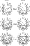
Results: This attempt to replicate previously published methods with this data set showed that patients with left-sided TLE had reduced connectivity between the posterior cingulate (PCC) and both the left (p = 0.012) and right (p < 0.002) hippocampus, while patients with right-sided TLE showed reduced connectivity between the PCC and right hippocampus (p < 0.004). After recoding ROIs by laterality, significantly diminished functional connectivity was observed between the PCC and hippocampus of both hemispheres (ipsilateral hippocampus, p < 0.001; contralateral hippocampus, p = 0.017) in patients with TLE compared with control participants. Regression analyses showed the reduced DMN recruitment of the ipsilateral hippocampus and parahippocampal gyrus (PHG) to be independent of clinical variables including hippocampal sclerosis, seizure frequency, and duration of illness. The graph theory metric of strength (or mean absolute correlation) showed significantly reduced connectivity of the ipsilateral hippocampus and ipsilateral PHG in patients with TLE compared with controls (hippocampus: p = 0.028; PHG: p = 0.021, after correction for false discovery rate). Finally, these hemispheric asymmetries in strength were observed in patients with TLE that corresponded to hemisphere of epileptogenic focus; 87% of patients with TLE had weaker ipsilateral hippocampus strength (compared with the contralateral hippocampus), and 80% of patients had weaker ipsilateral PHG strength.
Conclusions: This study demonstrated that recoding brain regions by the laterality to their epileptogenic focus increases the power of statistical approaches for finding interhemispheric differences in brain function. Using this approach, the authors showed TLE to selectively diminish connectivity of the hippocampus and parahippocampus in the hemisphere of the epileptogenic focus. This approach may prove to be a useful method for determining the seizure onset zone with TLE, and could be broadly applied to other neurological disorders with a lateralized onset.
Figures


Similar articles
-
Graph theory findings in the pathophysiology of temporal lobe epilepsy.Clin Neurophysiol. 2014 Jul;125(7):1295-305. doi: 10.1016/j.clinph.2014.04.004. Epub 2014 Apr 21. Clin Neurophysiol. 2014. PMID: 24831083 Free PMC article. Review.
-
Functional connectivity of hippocampal networks in temporal lobe epilepsy.Epilepsia. 2014 Jan;55(1):137-45. doi: 10.1111/epi.12476. Epub 2013 Dec 6. Epilepsia. 2014. PMID: 24313597 Free PMC article.
-
Graph theory application with functional connectivity to distinguish left from right temporal lobe epilepsy.Epilepsy Res. 2020 Nov;167:106449. doi: 10.1016/j.eplepsyres.2020.106449. Epub 2020 Sep 6. Epilepsy Res. 2020. PMID: 32937221
-
Functional connectivity of the hippocampus in temporal lobe epilepsy: feasibility of a task-regressed seed-based approach.Brain Connect. 2013;3(5):464-74. doi: 10.1089/brain.2013.0150. Brain Connect. 2013. PMID: 23869604 Free PMC article.
-
Mechanisms of cognitive impairment in temporal lobe epilepsy: A systematic review of resting-state functional connectivity studies.Epilepsy Behav. 2021 Feb;115:107686. doi: 10.1016/j.yebeh.2020.107686. Epub 2020 Dec 24. Epilepsy Behav. 2021. PMID: 33360743
Cited by
-
Hippocampal Atrophy Is Associated with Altered Hippocampus-Posterior Cingulate Cortex Connectivity in Mesial Temporal Lobe Epilepsy with Hippocampal Sclerosis.AJNR Am J Neuroradiol. 2017 Mar;38(3):626-632. doi: 10.3174/ajnr.A5039. Epub 2017 Jan 19. AJNR Am J Neuroradiol. 2017. PMID: 28104639 Free PMC article.
-
A functional connectivity-based neuromarker of sustained attention generalizes to predict recall in a reading task.Neuroimage. 2018 Feb 1;166:99-109. doi: 10.1016/j.neuroimage.2017.10.019. Epub 2017 Oct 12. Neuroimage. 2018. PMID: 29031531 Free PMC article.
-
Functional connectivity of hippocampus in temporal lobe epilepsy depends on hippocampal dominance: a systematic review of the literature.J Neurol. 2022 Jan;269(1):221-232. doi: 10.1007/s00415-020-10391-8. Epub 2021 Feb 10. J Neurol. 2022. PMID: 33564915
-
Voxel-wise Functional Connectivity of the Default Mode Network in Epilepsies: A Systematic Review and Meta-Analysis.Curr Neuropharmacol. 2022;20(1):254-266. doi: 10.2174/1570159X19666210325130624. Curr Neuropharmacol. 2022. PMID: 33823767 Free PMC article.
-
Graph theory findings in the pathophysiology of temporal lobe epilepsy.Clin Neurophysiol. 2014 Jul;125(7):1295-305. doi: 10.1016/j.clinph.2014.04.004. Epub 2014 Apr 21. Clin Neurophysiol. 2014. PMID: 24831083 Free PMC article. Review.
References
-
- Beck AT, Steer RA, Brown GK. Beck Depression Inventory - II. San Antonio, TX: Psychological Corp; 1996.
-
- Bettus G, Bartolomei F, Confort-Gouny S, Guedj E, Chauvel P, Cozzone PJ, et al. Role of resting state functional connectivity MRI in presurgical investigation of mesial temporal lobe epilepsy. J Neurol Neurosurg Psychiatry. 2010;81:1147–1154. - PubMed
-
- Buckner RL, Andrews-Hanna JR, Schacter DL. The brain’s default network: anatomy, function, and relevance to disease. Ann N Y Acad Sci. 2008;1124:1–38. - PubMed
Publication types
MeSH terms
Grants and funding
LinkOut - more resources
Full Text Sources
Other Literature Sources

