

Chest computed tomography performed on admission helps predict the severity of smoke-inhalation injury
- PMID: 23706091
- PMCID: PMC3707034
- DOI: 10.1186/cc12740
Chest computed tomography performed on admission helps predict the severity of smoke-inhalation injury
Abstract
Introduction: Smoke-inhalation injury is a major cause of mortality in burn patients, and therefore, it is important to determine accurately the severity of such injuries in these patients. The objective of this study was to evaluate whether chest computed tomography (CT) can be used for detecting early predictors of severity and complications of smoke-inhalation injury.
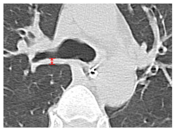
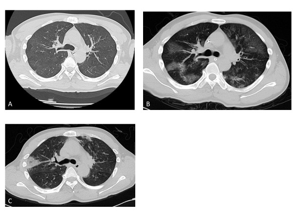
Methods: We evaluated 37 patients who had sustained smoke-inhalation injuries and had undergone chest CT within a few hours of admission to a hospital. Bronchoscopy was performed according to a standardized protocol within 12 hours of admission in all smoke-inhalation injury patients. Bronchial-wall thickness (BWT) was measured 2 cm distal from the tracheal bifurcation with CT images, and the following data were collected: total number of ventilator days, duration of intensive care unit (ICU) stay, pneumonia development, and patient outcome.
Results: The mean age of the patients was 63±18 years (range, 22 to 87 years), 31 (83.8%) of the patients were men, and the mortality rate was 10.8%. The causes of death in these patients were smoke inhalation (n=1), hemorrhage (n=1), and other factors resulting in sepsis (n=2). The initial carboxyhemoglobin level was 13%±14% (range, 1% to 50%). No significant correlation was found between bronchoscopic scoring and clinical factors. However, significant correlations were noted between admission BWT and development of pneumonia (R2=0.41; P<0.0001) and total number of ventilator days (R2=0.56; P<0.0001) and ICU-stay days (R2=0.17; P=0.01). Receiver operating characteristic curve analysis showed that an admission BWT cutoff value of >3.0 mm predicted pneumonia development with a sensitivity of 79%, specificity of 96%, positive predictive value of 91%, and negative predictive value of 88%.
Conclusion: BWT measured by using the chest CT scans obtained within a few hours of admission was predictive of the total number of ventilator days and ICU-stay days and the development of pneumonia in patients with smoke-inhalation injuries.
Figures
References
-
- Bingham HG, Gallagher TJ, Powell MD. Early bronchoscopy as a predictor of ventilatory support for burned patients. J Trauma. 1987;27:1286–1288. - PubMed
MeSH terms
LinkOut - more resources
Full Text Sources
Other Literature Sources
Medical
