

Variability in prostate and seminal vesicle delineations defined on magnetic resonance images, a multi-observer, -center and -sequence study
- PMID: 23706145
- PMCID: PMC3680182
- DOI: 10.1186/1748-717X-8-126
Variability in prostate and seminal vesicle delineations defined on magnetic resonance images, a multi-observer, -center and -sequence study
Abstract
Background: The use of magnetic resonance (MR) imaging as a part of preparation for radiotherapy is increasing. For delineation of the prostate several publications have shown decreased delineation variability using MR compared to computed tomography (CT). The purpose of the present work was to investigate the intra- and inter-physician delineation variability for prostate and seminal vesicles, and to investigate the influence of different MR sequence settings used clinically at the five centers participating in the study.
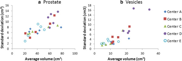
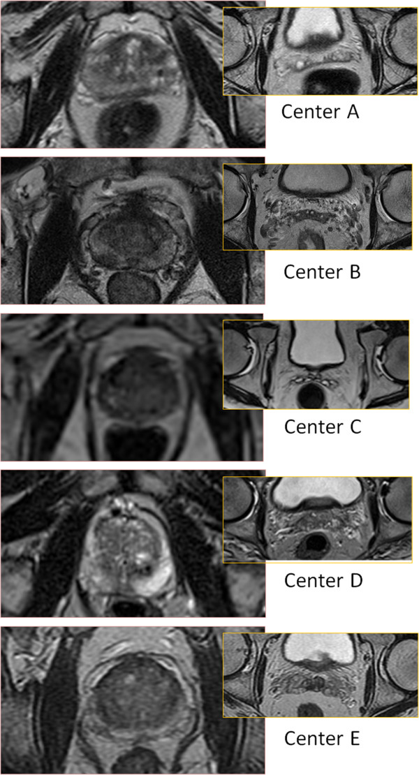
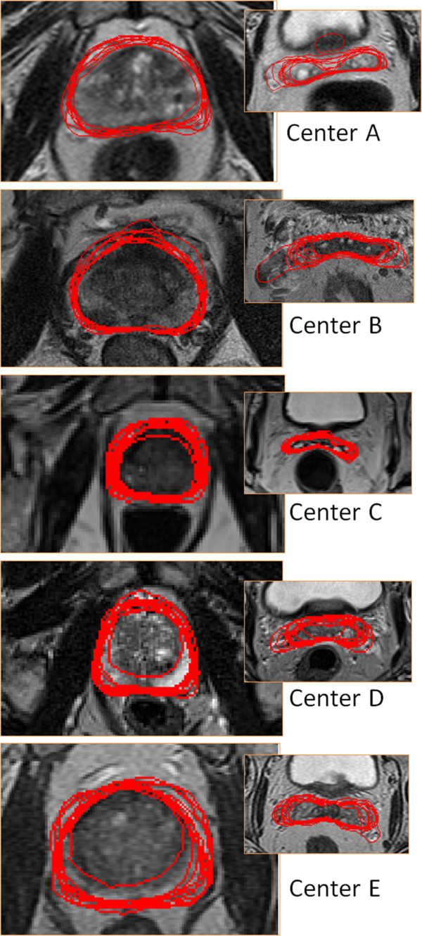
Methods: MR series from five centers, each providing five patients, were used. Two physicians from each center delineated the prostate and the seminal vesicles on each of the 25 image sets. The variability between the delineations was analyzed with respect to overall, intra- and inter-physician variability, and dependence between variability and origin of the MR images, i.e. the MR sequence used to acquire the data.
Results: The intra-physician variability in different directions was between 1.3 - 1.9 mm and 3 - 4 mm for the prostate and seminal vesicles respectively (1 std). The inter-physician variability for different directions were between 0.7 - 1.7 mm and approximately equal for the prostate and seminal vesicles. Large differences in variability were observed for individual patients, and also for individual imaging sequences used at the different centers. There was however no indication of decreased variability with higher field strength.
Conclusion: The overall delineation variability is larger for the seminal vesicles compared to the prostate, due to a larger intra-physician variability. The imaging sequence appears to have a large influence on the variability, even for different variants of the T2-weighted spin-echo based sequences, which were used by all centers in the study.
Figures


References
-
- Khoo VS, Joon DL. New developments in MRI for target volume delineation in radiotherapy. Br J Radiol. 2006;79 Spec No:S2–S15. - PubMed
Publication types
MeSH terms
LinkOut - more resources
Full Text Sources
Other Literature Sources
Medical
Research Materials
