. 2013 May 24;15(1):42.
doi: 10.1186/1532-429X-15-42.

SHILO, a novel dual imaging approach for simultaneous HI-/LOw temporal (Low-/Hi-spatial) resolution imaging for vascular dynamic contrast enhanced cardiovascular magnetic resonance: numerical simulations and feasibility in the carotid arteries

Affiliations

SHILO, a novel dual imaging approach for simultaneous HI-/LOw temporal (Low-/Hi-spatial) resolution imaging for vascular dynamic contrast enhanced cardiovascular magnetic resonance: numerical simulations and feasibility in the carotid arteries

Claudia Calcagno et al. J Cardiovasc Magn Reson. .

Abstract

Background: Dynamic contrast enhanced (DCE) cardiovascular magnetic resonance (CMR) is increasingly used to quantify microvessels and permeability in atherosclerosis. Accurate quantification depends on reliable sampling of both vessel wall (VW) uptake and contrast agent dynamic in the blood plasma (the so called arterial input function, AIF). This poses specific challenges in terms of spatial/temporal resolution and matched dynamic MR signal range, which are suboptimal in current vascular DCE-CMR protocols. In this study we describe a novel dual-imaging approach, which allows acquiring simultaneously AIF and VW images using different spatial/temporal resolution and optimizes imaging parameters for the two compartments. We refer to this new acquisition as SHILO, Simultaneous HI-/LOw-temporal (low-/hi-spatial) resolution DCE-imaging.

Methods: In SHILO, the acquisition of low spatial resolution single-shot AIF images is interleaved with segments of higher spatial resolution images of the VW. This allows sampling the AIF and VW with different spatial/temporal resolution and acquisition parameters, at independent spatial locations. We show the adequacy of this temporal sampling scheme by using numerical simulations. Following, we validate the MR signal of SHILO against a standard 2D spoiled gradient recalled echo (SPGR) acquisition with in vitro and in vivo experiments. Finally, we show feasibility of using SHILO imaging in subjects with carotid atherosclerosis.

Results: Our simulations confirmed the superiority of the SHILO temporal sampling scheme over conventional strategies that sample AIF and tissue curves at the same time resolution. Both the median relative errors and standard deviation of absolute parameter values were lower for the SHILO than for conventional sampling schemes. We showed equivalency of the SHILO signal and conventional 2D SPGR imaging, using both in vitro phantom experiments (R2 =0.99) and in vivo acquisitions (R2 =0.95). Finally, we showed feasibility of using the newly developed SHILO sequence to acquire DCE-CMR data in subjects with carotid atherosclerosis to calculate plaque perfusion indices.

Conclusions: We successfully demonstrate the feasibility of using the newly developed SHILO dual-imaging technique for simultaneous AIF and VW imaging in DCE-CMR of atherosclerosis. Our initial results are promising and warrant further investigation of this technique in wider studies measuring kinetic parameters of plaque neovascularization with validation against gold standard techniques.


Figures

Figure 1
Figure 1
Schematic representation of the SHILO segmentation/interleaving scheme. The diagram illustrates an example where the high spatial resolution image is acquired in 4 segments, thus leading to the acquisition of 4 single shot AIF images for each segmented high resolution image. L1-4, single shot AIF images. A-D, segments of the multi-shot tissue image. H1: multi-shot high spatial resolution image coming from the combination of segments A-D. Low spatial resolution images L1-4 are acquired in location L, and high spatial resolution image H1 is acquired in independent location H. The black bar represents the saturation pulse preceding the low spatial resolution AIF image and high spatial resolution segment.
Figure 2
Figure 2
Flow chart of in vivo CMR experiment. After anatomical black blood imaging a low dose (0.01 mmol/Kg) 2D SPGR and a low dose (0.01 mmol/Kg) SHILO scans are acquired for in vivo validation of the SHILO MR signal. Following, a higher dose (0.05 mmol/Kg) SHILO acquisition was used to calculate perfusion parameters in the carotid vessel wall and sternocleidomastoid skeletal muscle.
Figure 3
Figure 3
Effect of under-sampling on simulated AIF (panel A) and three simulated tissue curves (panels B to D). Different color curves represent different degrees of under-sampling, corresponding to decreasing time resolutions for an increasing number of imaged tissue slices, as detailed in the legend. Kinetic parameters used are specified in the legends.
Figure 4
Figure 4
Results of numerical simulations for percentage error. Panel A, vp. Panel B, Ktrans. Panel C, ve. Panel D, Kep. Red dashed line, results for the same time resolution (STR) sampling scheme. Green solid line, results for the different time resolution (DTR) sampling scheme implemented in SHILO (AIF time resolution 1.6 s). Blue dash dotted line, results for the second different time resolution (DTR) sampling scheme with AIF time resolution 0.8 s.
Figure 5
Figure 5
Results of numerical simulations for percentage standard deviation. Panel A, vp. Panel B, Ktrans. Panel C, ve. Panel D, Kep. Red dashed line, results for the same time resolution (STR) sampling scheme. Green solid line, results for the different time resolution (DTR) sampling scheme implemented in SHILO (AIF time resolution 1.6 s). Blue dash dotted line, results for the second different time resolution (DTR) sampling scheme with AIF time resolution 0.8 s.
Figure 6
Figure 6
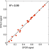
Correlation of normalized signal values from SHILO and equivalent SPGR acquisitions in a phantom with a range of T1 values corresponding to gadolinium concentrations between 0 and 5 mM. Correlation coefficient R2 = 0.99.
Figure 7
Figure 7
AIF concentration-time curves measured using SHILO and an equivalent SPGR acquisition. Panel A shows the average AIF for all vessels in all subjects. Panel B shows the AIF from one vessel in one subject. Panel C shows the correlation between the in vivo average SPGR and SHILO signals (R2 = 0.95; slope 0.95, intercept - 0.01).
Figure 8
Figure 8
Anatomical images of a representative subject, one axial slice and zoomed in insert on the right common carotid artery (bottom left of all panels). Panel A, SHILO low spatial/high temporal resolution image for AIF acquisition. Red star in insert, ROI used to sample blood SI curve. Panel B, SHILO high spatial/low temporal resolution image for tissue data acquisition. In the insert, the ROIs used to sample the muscle (green) and vessel wall (blue) SI curves are depicted. Panel C: red line, AIF from low spatial/high temporal resolution SHILO; blue dots, vessel wall SI curve from high spatial/low temporal resolution SHILO; green dots, muscle SI curve from high spatial/low temporal resolution SHILO. Blue and green lines represent kinetic modeling fittings. X axis, time (min). Y axis, contrast agent concentration (mmol/l). Vessel wall and muscle curves are plotted 25 times their measured concentrations for clarity in the plot.

References

    1. Moreno PR, Purushothaman KR, Fuster V, Echeverri D, Truszczynska H, Sharma SK, Badimon JJ, O’Connor WN. Plaque neovascularization is increased in ruptured atherosclerotic lesions of human aorta: implications for plaque vulnerability. Circulation. 2004;110:2032–8. doi: 10.1161/01.CIR.0000143233.87854.23. - DOI - PubMed
    1. Choyke PL, Dwyer AJ, Knopp MV. Functional tumor imaging with dynamic contrast-enhanced magnetic resonance imaging. J Magn Reson Imaging. 2003;17:509–20. doi: 10.1002/jmri.10304. - DOI - PubMed
    1. Notohamiprodjo M, Reiser MF, Sourbron SP. Diffusion and perfusion of the kidney. Eur J Radiol. 2010;76:337–47. doi: 10.1016/j.ejrad.2010.05.033. - DOI - PubMed
    1. Padhani AR. Dynamic contrast-enhanced MRI in clinical oncology: current status and future directions. J Magn Reson Imaging. 2002;16:407–22. doi: 10.1002/jmri.10176. - DOI - PubMed
    1. Calcagno C, Cornily JC, Hyafil F, Rudd JH, Briley-Saebo KC, Mani V, Goldschlager G, Machac J, Fuster V, Fayad ZA. Detection of neovessels in atherosclerotic plaques of rabbits using dynamic contrast enhanced MRI and 18F-FDG PET. Arterioscler Thromb Vasc Biol. 2008;28:1311–7. doi: 10.1161/ATVBAHA.108.166173. - DOI - PMC - PubMed

Publication types