Review
. 2013 Aug;23(4):461-9.
doi: 10.1016/j.gde.2013.04.001. Epub 2013 May 21.

Genetic dissection of ventral folding morphogenesis in mouse: embryonic visceral endoderm-supplied BMP2 positions head and heart

Affiliations
Review

Genetic dissection of ventral folding morphogenesis in mouse: embryonic visceral endoderm-supplied BMP2 positions head and heart

Svetlana Gavrilov et al. Curr Opin Genet Dev. 2013 Aug.

Abstract

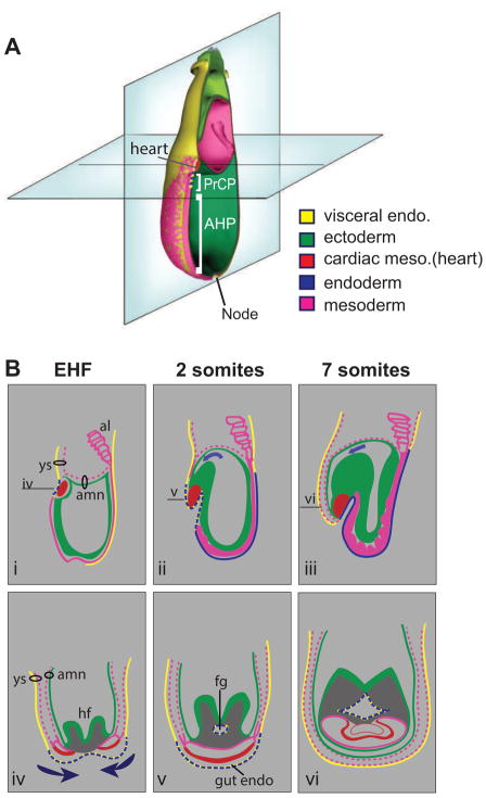
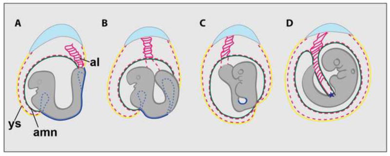
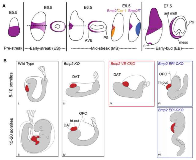
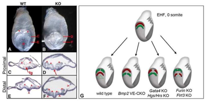
Ventral folding morphogenesis (VFM), a vital morphogenetic process in amniotes, mediates gut endoderm internalization, linear heart tube formation, ventral body wall closure and encasement of the fetus in extraembryonic membranes. Aberrant VFM underlies a number of birth defects such as gastroschisis and ectopia cordis in human and misplacement of head and heart in mouse. Recent cell lineage-specific mouse mutant analyses identified the Bone Morphogenetic Protein (BMP) pathway and Anterior Visceral Endoderm (AVE) as key regulators of anterior VFM. Loss of BMP2 expression solely from embryonic visceral endoderm (EmVE) and the AVE blocks formation of foregut invagination, and simultaneously, aberrantly positions the heart anterior/dorsal to the head, suggesting a mechanistic link between foregut and head/heart morphogenesis.


Figures

Figure 1
Figure 1. Ventral folding morphogenesis encompasses multiple concurrent tissue rearrangements: EHF, 0-to-8 somites
(A) An oblique frontal view of an EHF mouse embryo; planes indicate positions of sagittal and transverse sections shown in B. Before the onset of VFM, ectoderm (green) lines the amniotic cavity and encompasses the inner-most layer of the distal (embryonic) portion of the conceptus; endoderm (yellow, blue) forms the surface layer and overlies the mesoderm (pink). The heart primordia (red), which develop bilaterally from splanchnic mesoderm, reside just anterior to the nascent ectodermal headfolds [6]. The anterior midline consists of three structures [9,11]: the node, which will generate trunk and tail notochord; anterior head process (AHP), axial mesoderm -derived from the early/mid gastrula organizer- that underlies the prospective midbrain and rostral hindbrain; and Prechordal plate (PrCP), axial mesendoderm -derived from the early gastrula organizer- underlies the prospective forebrain. (B) The tissue folding propelling VFM occurs along two axes, rostral-caudal (depicted in sagittal sections i–iii) and ventral-to-lateral (depicted in transverse sections, iv–vi), during the EHF (i, iv), 2 somites (ii, v), and 7 somites (iii, vi) stages. The horizontal lines (labeled iv, v, vi) in respectively, panels i, ii, and iii indicate the level of the transverse section in the corresponding lower panel. Rostral-caudal folding repositions endoderm cells along the midline as it transforms the sheet of anterior gut endoderm from a convex external layer into a concave internal foregut pocket protruding rostrally [–8]. Concurrent rostral-caudal folding of neuroectoderm repositions the headfolds to reside anterior and dorsal to cardiac mesoderm [4,6,39]. Lateral-to-ventral folding of the bilateral heart primordia positions cardiac progenitors at the ventral line, where they merge to form the primitive heart tube. Concomitantly, lateral regions of gut endoderm and of the body wall (apposed somatic mesoderm and surface ectoderm layers) move ventrally toward the midline and fuse to close, respectively, the foregut tube and the body wall. The visceral yolk sac (extraembryonic visceral endoderm –yellow plus extraembryonic mesoderm –pink dashed line) undergoes ventral-to-lateral tissue movements to envelope the anterior region (head and heart) of the early-somite embryo. Arrows indicate direction of rostral-caudal (ii, iii) and lateral-to-ventral (iv) folding. Abbreviations: AHP- anterior head process; amn- amnion; fg, foregut pocket; hf-head folds; PrCP-prechordal plate; ys-visceral yolk sac.
Figure 2
Figure 2. Ventral folding morphogenesis encompasses multiple concurrent tissue rearrangements: 8-to-20 somites
(A) 8–10 somites; (B) 10–12 somites; (C) 12–14 somites; (D) 15–20 somites. (A) 8–10 somite stage embryo at the onset of turning with hindgut and foregut pockets (dashed blue lines), external midgut region (solid blue line), and the ventral region, caudal to the heart, outside the amnion and yolk sac. Lateral edges of the amnion and yolk sac are joined to the embryo at the umbilical ring (boundary between the body wall and future attachment site of the umbilical cord, an allantois derivative). The amniotic ectoderm (green line) exists as an extension of the embryonic ectoderm, whilst the amniotic mesoderm lies in direct contact with the mesodermal layer of the visceral yolk sac (red dashed lines). (B–D) Turning/axial rotation inverts the lordotic U-shaped trunk region, placing the embryo in the fetal position with the head looking toward, rather than away from, the tail region; (compare A and D). Upon completion of turning, with embryo now encased within the extraembryonic membranes (amnion and visceral yolk sac), the ectoderm and mesoderm layers of the ventral body wall are continuous with the ectoderm and mesoderm layers of the amnion, which now covers the surface of the allantois/umbilical cord (D) [43]. Turning places the umbilical cord in the midline, at the meeting point of the original anterior and posterior connection sites between the amnion and embryo (* in D). al- allantois; amn- amnion; ys- visceral yolk sac.
Figure 3
Figure 3. BMP2 expression in EmVE/AVE and epiblast lineages regulates distinct steps in VFM
(A) Schematic diagrams of Bmp2 expression in EmVE during the pre-streak (E5.5), early-streak (E6.5); mid-streak (E6.5) and early bud (E7.5) stages. Diagrams illustrate lateral views or transverse sections at the indicated levels; anterior is to the left. Results of two-color in situ hybridizations to Bmp2 (purple) and Cer1 (orange) or Brachyury/T (blue) are illustrated. ant midl- anterior midline; AVE- anterior visceral endoderm; EB-early bud; ES- early streak; MS-mid- streak; meso-mesoderm; n-node; PS- primitive streak. (B) Schematic representations of lineage specific Bmp2 knockout phenotypes. (i and ii) Wild type embryos show proper arrangement of the head, heart, amnion (thin gray line) and allantois (hatched gray lines) before (8–10 somites) and after turning (15–20 somites). The heart lays posterior/ventral to the head (8–10 somites) and has undergone looping (15–20 somites); both head and heart sit inside the amnion. Bmp2 KO embryos display one or more of four defects: (iii) disorganized anterior phenotype (DAT); (iv) open proamniotic canal (OPC), heart outside the amnion (ht-out), DAT and unturned. (v) Bmp2 VE-CKO displays DAT phenotype; (vi) Bmp2 EPI-CKO displays ht-out and OPC phenotype (8–10 somites) plus (vii) posterior specific delay (15–20 somites) in which the heart fails to loop and the embryo remains unturned.
Figure 4
Figure 4. Disorganized anterior phenotype: formation of foregut invagination and linear heart tube are independent morphogenetic events
(A–F) Bmp2 KO mutants with the disorganized anterior phenotype display ectopic neural folds and absence of foregut invagination. Frontal views of (A) WT and (B) KO embryos at the 4 somite stage. The two symmetric head folds (red asterisks) and a single neural groove (red ^) present in WT are absent in the KO. (C–E) Transverse sections of WT at the regions indicated in A; (D–F) transverse sections of KO at the regions indicated in B; anterior/ventral is downward. (C) Proximal WT section showing symmetrical head folds and foregut. (E) Distal WT section showing two head folds, medial hinge point (red ^) and surface ectoderm (red >). (D) Proximal KO section showing uneven head folds (red asterisks) and absence of the foregut pocket (red arrow). (F) Distal KO section showing additional neural folds (red asterisks), ectopic hinge points (red ^), and surface ectoderm (red >); (reproduced with permission from Madabhushi and Lacy [23**]). (G) Model for origin of cardia bifida. EHF (0 somite) embryo before onset of VFM is shown on top; the heart progenitors (red) reside just anterior to the nascent ectodermal headfolds (green). Arrows point to four different scenarios at the 8–10 somite stage: 1) wild type - rostral-caudal folding positions the heart posterior to the head folds and generates the foregut invagination; lateral-to-ventral folding fuses the bilateral heart primordia and closes the gut tube at the ventral midline (closed yellow-blue triangle); 2) Bmp2 VE-CKO – absence of rostral-caudal folding results in absence of foregut invagination and retention of heart progenitors anterior to the headfolds; the presence of a single heart field indicates that lateral-to-ventral folding occurred to fuse the bilateral heart primordia; 3) Gata4 KO or Hrs KO – similar to the Bmp2 VE-CKO absence of rostral-caudal folding results in absence of foregut invagination and retention of heart progenitors anterior to the headfolds; but lateral-to-ventral folding also does not occur, resulting in lack of fusion of the anteriorly mispositioned bilateral heart primordia; and 4) Furin KO or Flrt3 KO - rostral-caudal folding occurs and positions the heart primordia posterior to the head folds and generates the foregut invagination but absence of lateral-to-ventral folding results in lack of fusion of the properly posteriorly positioned bilateral heart primordia and failure to close the foregut tube at the ventral midline (open yellow-blue triangle). The diagrams are based on Madabhushi and Lacy [23**] and on targeted mutations in the transcription factor Gata4 [–38*], the vesicular trafficking protein Hgs/Hrs (HGF-regulated tyrosine kinase substrate) [39*], Furin, a proprotein convertase [40, 42*], and Flrt3 (fibronectin leucine rich transmembrane protein 3) [41*].

References

    1. Sadler TW, Feldkamp ML. The embryology of body wall closure: relevance to gastroschisis and other ventral body wall defects. Am J Med Genet C Semin Med Genet. 2008;148C:180–185. - PubMed
    1. Williams T. Animal models of ventral body wall closure defects: a personal perspective on gastroschisis. Am J Med Genet C Semin Med Genet. 2008;148C:186–191. - PubMed
    1. Frolov P, Alali J, Klein MD. Clinical risk factors for gastroschisis and omphalocele in humans: a review of the literature. Pediatr Surg Int. 2010;26:1135–1148. - PubMed
    1. Kaufman MB. The Atlas of Mouse Development. London: Academic Press; 1992.
    1. Sadler TW. The embryologic origin of ventral body wall defects. Semin Pediatr Surg. 2010;19:209–214. - PubMed

Publication types

Substances

LinkOut - more resources