. 2013 May 23;38(5):970-83.
doi: 10.1016/j.immuni.2013.04.011.

IRF4 transcription factor-dependent CD11b+ dendritic cells in human and mouse control mucosal IL-17 cytokine responses

Affiliations

IRF4 transcription factor-dependent CD11b+ dendritic cells in human and mouse control mucosal IL-17 cytokine responses

Andreas Schlitzer et al. Immunity. .

Abstract

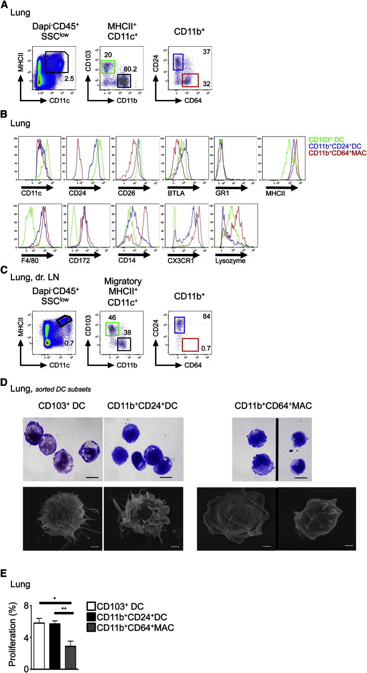
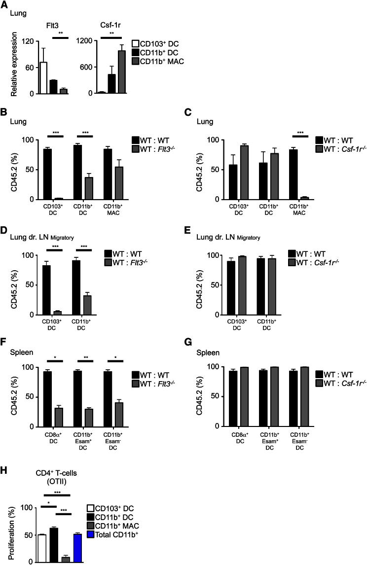
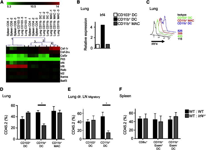
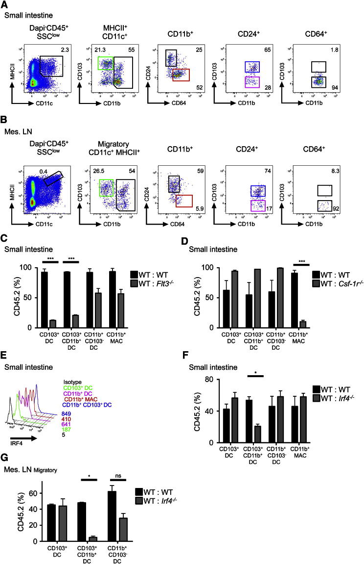
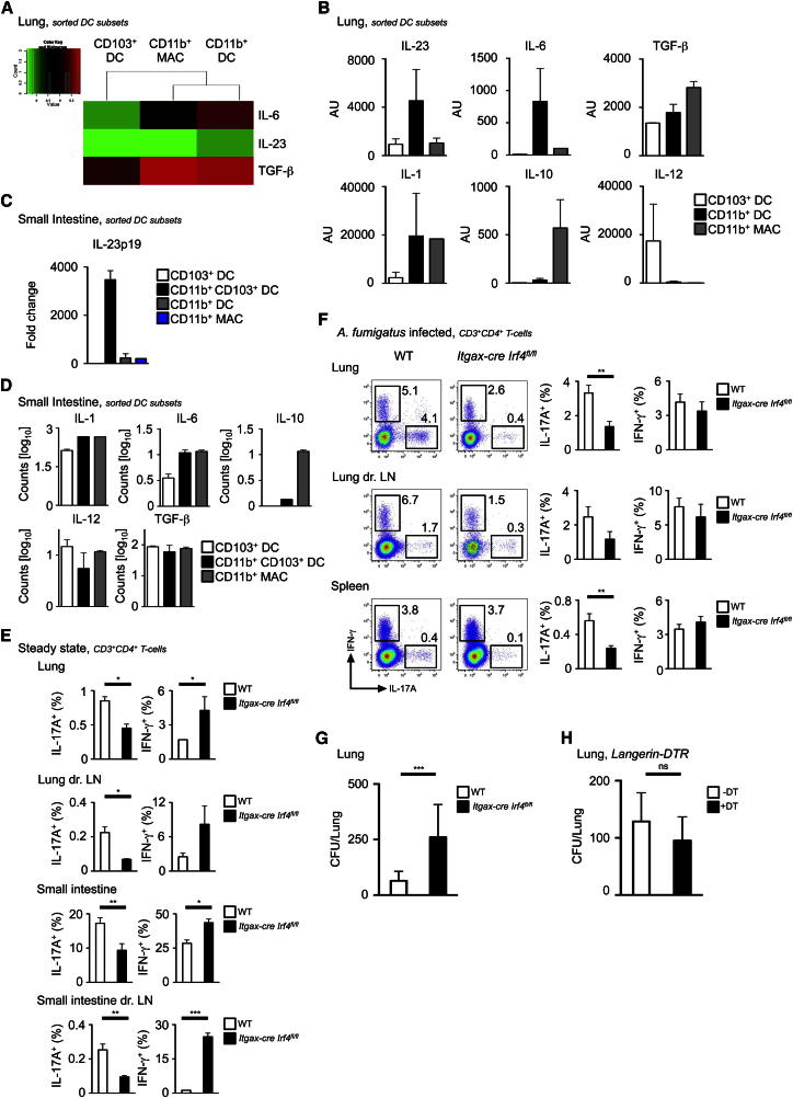
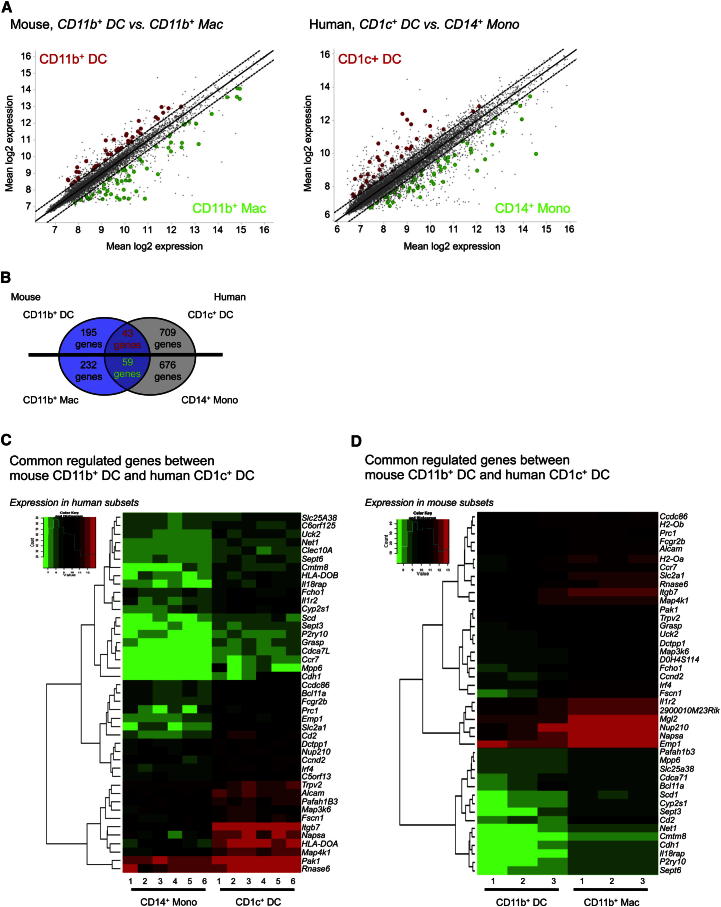
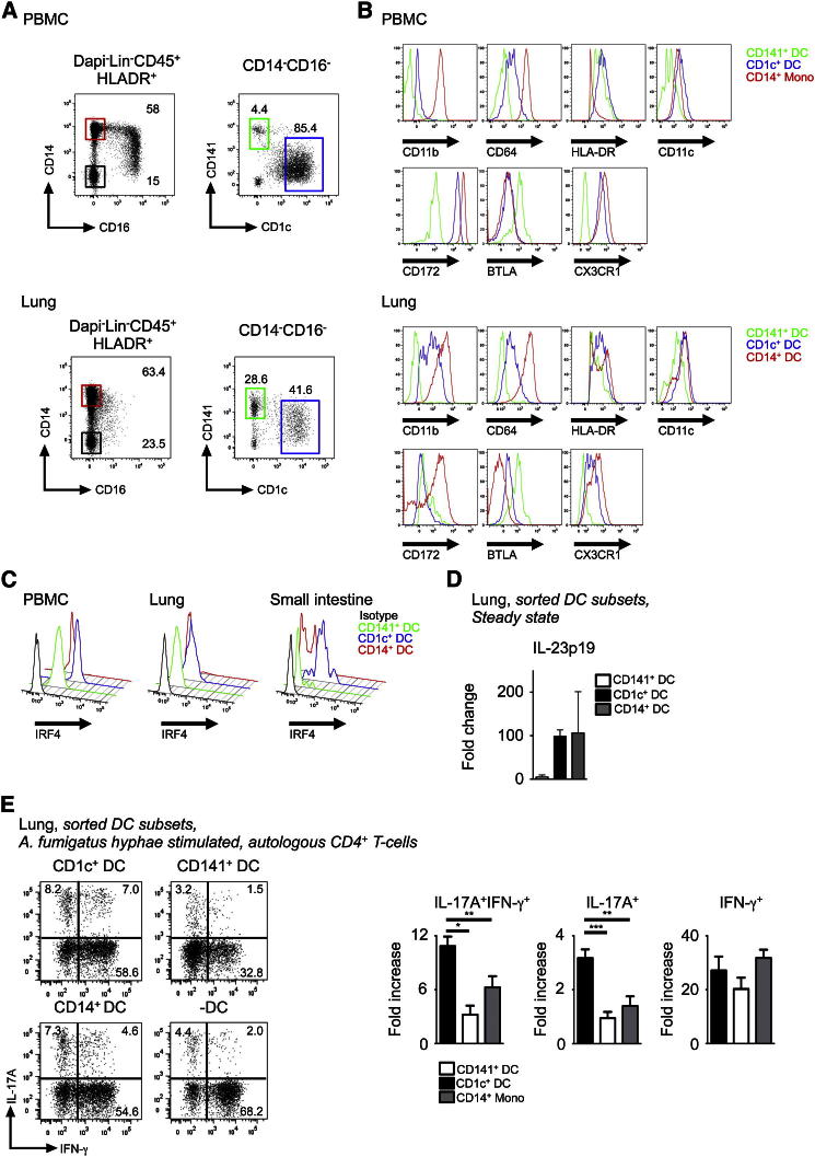
Mouse and human dendritic cells (DCs) are composed of functionally specialized subsets, but precise interspecies correlation is currently incomplete. Here, we showed that murine lung and gut lamina propria CD11b+ DC populations were comprised of two subsets: FLT3- and IRF4-dependent CD24(+)CD64(-) DCs and contaminating CSF-1R-dependent CD24(-)CD64(+) macrophages. Functionally, loss of CD24(+)CD11b(+) DCs abrogated CD4+ T cell-mediated interleukin-17 (IL-17) production in steady state and after Aspergillus fumigatus challenge. Human CD1c+ DCs, the equivalent of murine CD24(+)CD11b(+) DCs, also expressed IRF4, secreted IL-23, and promoted T helper 17 cell responses. Our data revealed heterogeneity in the mouse CD11b+ DC compartment and identifed mucosal tissues IRF4-expressing DCs specialized in instructing IL-17 responses in both mouse and human. The demonstration of mouse and human DC subsets specialized in driving IL-17 responses highlights the conservation of key immune functions across species and will facilitate the translation of mouse in vivo findings to advance DC-based clinical therapies.


Figures

Figure 1
Figure 1
Lung CD11b+ DCs Are Heterogeneous (A) Flow cytometry of mouse lung cell suspension. Gating strategy to identify CD103+ DCs (green gate), CD11b+CD24+ DCs (blue gate), and CD11b+CD64+ MACs (red gate) is shown. (B and C) Histograms show relative expression of the indicated markers on lung DCs and MACs (B). Flow cytometry of mouse LLN cell suspension. Gating strategy as described in (A) was used in (C). Representative data from n > 5 shown for (A)–(C). (D) Morphology of purified lung DCs and MACs visualized by GIEMSA staining and SEM. (E) Percentage proliferation of lung DCs and MACs indicated by fluorescence levels in Fucci mice (mean fluorescent+ cells ± SEM, pooled results of three experiments, n = 6). See also Figure S1.
Figure 2
Figure 2
Lung CD11b+CD24+CD64 Cells Are Bona Fide DCs, whereas CD11b+CD24CD64+ Cells Are Macrophages (A) Relative expression of Flt3 and Csf-1r mRNA by lung DCs and MACs (n = 3, mean ± SEM). (B–G) Percentage of CD45.2 and CD45.1 DCs and MACs in the indicated tissues from mixed BM chimeric mice (CD45.2 WT: CD45.1 WT; CD45.2 Flt3−/−: CD45.1 WT (B, D, F); CD45.2 Csf1r−/−: CD45.1 WT (C, E, G). (H) Bar graph shows mean ± SEM of the indicated cell populations (pooled results of four experiments, n = 12). Percentage proliferation of OTII cells stimulated by OVA-loaded lung DCs and MACs (n = 3, mean ± SEM). See also Figure S2.
Figure 3
Figure 3
Lung CD11b+ DCs Are IRF4 Dependent (A) Hierarchical clustering of murine spleen and lung DC/MAC populations based on differential expression of selected genes (SAM algorithm, delta = 2.6). (B) Irf4 mRNA expression by lung DCs and MACs (n = 3, mean). (C) Relative expression of IRF4 by flow cytometry on lung CD11b+ DCs and MACs (n = 3). (D–F) Percentage of CD45.2 and CD45.1 DCs and MACs in the indicated tissues from indicated mixed BM chimeric mice. Bar graphs show mean ± SEM of the indicated cell populations (pooled results of four experiments, n = 10, mean ± SEM). See also Figure S2 and Table S1.
Figure 4
Figure 4
Gut Lamina Propria CD24+CD103+CD11b+ DCs Are Dependent on IRF4 (A) Flow cytometry of gut LP to identify CD103+ DCs, CD103+CD11b+ DCs, CD103-CD11b+ DCs, and CD11b+ MACs (n = 5). (B) Flow cytometry of MLN to identify DCs and MACs as described in (A), (n = 5). (C, D, F, and G) Percentage of CD45.2 and CD45.1 DCs and MACs in the indicated tissues from indicated BM chimeric mice. (E) Pooled results from four experiments, n = 10 (mean ± SEM). IRF4 expression on SI-LP DCs and MACs by flow cytometry (n = 3). See also Figure S3.
Figure 5
Figure 5
CD11b+CD24+ DCs in Lung and SI Control Th17 Responses (A–D) Expression of indicated genes in lung DCs and MACs by microarray (A), by Q-PCR in lung (B), and Q-PCR and nanostring in SI-LP (C and D) DCs and MACs (mean ± SEM, n = 3). (E) Percentage of IL-17A and IFN-γ expressing CD3+CD4+ T cells in steady state from lung, lymph node, SI-LP, and MLN of WT (white bars) and Itgax-cre Irf4fl/fl (black bars) mice (mean ± SEM, pooled results of two experiments, n = 10). (F) Same as (E) after A. fumigatus challenge. (F) Representative dot plot shown left and bar graphs show composite data (mean ± SEM, pooled results of four experiments, n = 20). (G) CFUs from homogenized lung tissue of either WT (white bars) or Itgax-cre Irf4fl/fl (black bars) mice 7 days after intranasal infection with A. fumigatus (mean ± SEM, pooled results of four experiments, n = 20). (H) Same than (F) with Langerin-DTR mice (white bars) or Langerin-DTR mice injected with DT (black bars) (mean ± SEM, pooled results of two experiments, n = 10). See also Figures S4 and S5.
Figure 6
Figure 6
Transcriptomic Alignment of Mouse IRF4-Dependent CD11b+ DCs with Human CD1c+ DCs (A) Scatterplot of mouse CD11b+ DCs versus CD11b+ MACs and human CD1c+ DCs and CD14+ monocytes. Common DEG for mouse CD11b+ DCs and human CD1c+ DCs are highlighted in red, whereas common DEG for mouse CD11b+ MACs and CD14+ monocytes are highlighted in green. All genes depicted are regulated < 1.5-fold. (B) Analysis strategy depicting the number of DEG, which are shared and not shared between mouse CD11b+ DCs, mouse CD11b+ MACs, human CD1c+ DCs, and human CD14+ monocytes. (C) Heatmap showing expression in CD14+ monocytes and CD1c+ DCs of common DEG identified between mouse CD11b+ DCs and human CD1c+ DCs. (D) Same than (C) with mouse CD11b+ DCs and CD11b+ MACs. See also Figure S7 and Tables S2 and S3.
Figure 7
Figure 7
Human IRF4 Expressing CD1c+ DCs Induce IL-17 T Helper Response Flow cytometry of peripheral blood and mechanically dispersed lung. Gating strategy used to identify three myeloid DC subsets within LinHLA-DR+ fraction (CD123+ pDCs were excluded from the CD14CD16 fraction): (1) CD14+ monocytes (red gate), (2) CD14CD1c+CD141 DCs (blue gate), (3) CD14CD1cCD141+ DCs (green gate). (A) Representative from ten blood and three lung donors are shown. Relative expression of indicated markers by CD14+ monocytes/DCs (red), CD1c+ DCs (blue), and CD141+ DCs (green). (B) Representative data from three blood and lung donors are shown. (C) Same as (B) for IRF4 expression compared to isotype (black) from indicated tissues. Representative data from three blood, lung, and two SI donors are shown. mRNA expression of IL-23p19 by freshly sorted human DC subsets from lung (n = 3, mean ± SEM). (D) Expression normalized to IL-23p19 mRNA expression in unstimulated CD141+ DCs. Intracellular IFN-γ and IL-17A expression by PMA/Ionomycin restimulated CD4+ T cells cultured with autologous lung indicated DC subsets pulsed with A. fumigatus hyphae. (E) Representative dot plot with percentage of total IL-17+ cells and composite results shown in bar graph (n = 4, mean ± SEM fold increase [%+DC/%-DC]).

References

    1. Acosta-Rodriguez E.V., Rivino L., Geginat J., Jarrossay D., Gattorno M., Lanzavecchia A., Sallusto F., Napolitani G. Surface phenotype and antigenic specificity of human interleukin 17-producing T helper memory cells. Nat. Immunol. 2007;8:639–646. - PubMed
    1. Aggarwal S., Ghilardi N., Xie M.H., de Sauvage F.J., Gurney A.L. Interleukin-23 promotes a distinct CD4 T cell activation state characterized by the production of interleukin-17. J. Biol. Chem. 2003;278:1910–1914. - PubMed
    1. Ahern P.P., Schiering C., Buonocore S., McGeachy M.J., Cua D.J., Maloy K.J., Powrie F. Interleukin-23 drives intestinal inflammation through direct activity on T cells. Immunity. 2010;33:279–288. - PMC - PubMed
    1. Annacker O., Coombes J.L., Malmstrom V., Uhlig H.H., Bourne T., Johansson-Lindbom B., Agace W.W., Parker C.M., Powrie F. Essential role for CD103 in the T cell-mediated regulation of experimental colitis. J. Exp. Med. 2005;202:1051–1061. - PMC - PubMed
    1. Bachem A., Güttler S., Hartung E., Ebstein F., Schaefer M., Tannert A., Salama A., Movassaghi K., Opitz C., Mages H.W. Superior antigen cross-presentation and XCR1 expression define human CD11c+CD141+ cells as homologues of mouse CD8+ dendritic cells. J. Exp. Med. 2010;207:1273–1281. - PMC - PubMed

Publication types

MeSH terms

Associated data