. 2013 May 23;153(5):1012-24.
doi: 10.1016/j.cell.2013.04.032.

Acetylation-mediated proteasomal degradation of core histones during DNA repair and spermatogenesis

Affiliations

Acetylation-mediated proteasomal degradation of core histones during DNA repair and spermatogenesis

Min-Xian Qian et al. Cell. .

Abstract

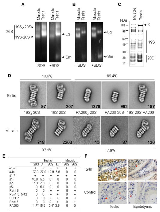
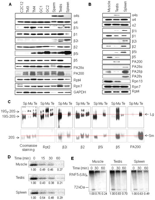
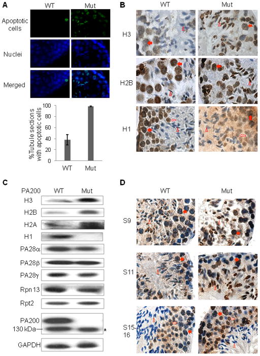
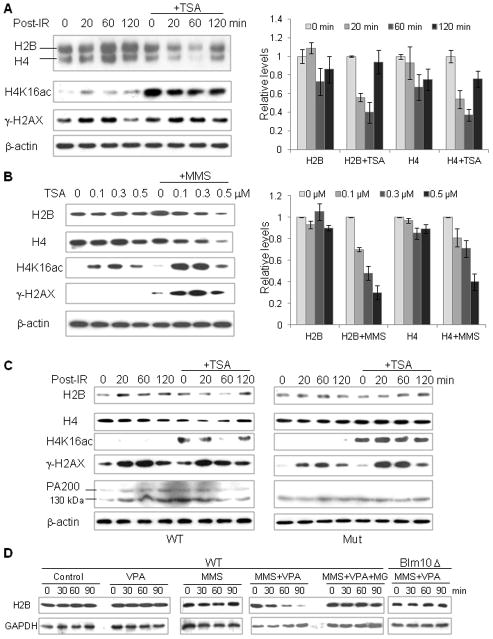
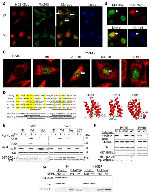
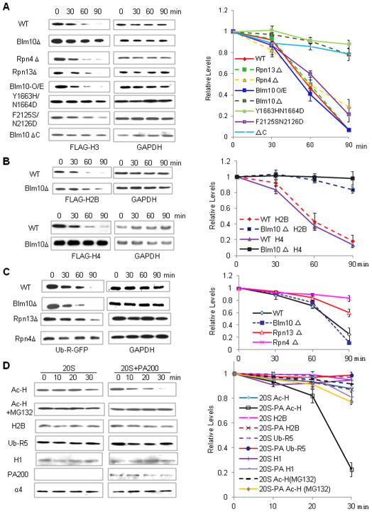
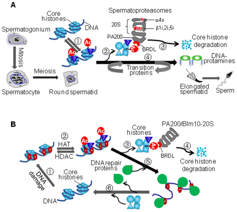
Histone acetylation plays critical roles in chromatin remodeling, DNA repair, and epigenetic regulation of gene expression, but the underlying mechanisms are unclear. Proteasomes usually catalyze ATP- and polyubiquitin-dependent proteolysis. Here, we show that the proteasomes containing the activator PA200 catalyze the polyubiquitin-independent degradation of histones. Most proteasomes in mammalian testes ("spermatoproteasomes") contain a spermatid/sperm-specific α subunit α4 s/PSMA8 and/or the catalytic β subunits of immunoproteasomes in addition to PA200. Deletion of PA200 in mice abolishes acetylation-dependent degradation of somatic core histones during DNA double-strand breaks and delays core histone disappearance in elongated spermatids. Purified PA200 greatly promotes ATP-independent proteasomal degradation of the acetylated core histones, but not polyubiquitinated proteins. Furthermore, acetylation on histones is required for their binding to the bromodomain-like regions in PA200 and its yeast ortholog, Blm10. Thus, PA200/Blm10 specifically targets the core histones for acetylation-mediated degradation by proteasomes, providing mechanisms by which acetylation regulates histone degradation, DNA repair, and spermatogenesis.


Figures

Figure 1
Figure 1. Identification of Two Distinct Types of Proteasomes in Mammalian Testes
(A) The proteasomes in bovine testes migrate differently from those in muscle on native PAGE. Proteasomes in crude tissue extracts were detected by incubating the gel with LLVY-amc in the absence or presence of 0.02% SDS, and visualized under UV light. Two unusual bands were labeled as Lg and Sm, respectively. (B) Proteasomes purified from seminiferous tubules of the testis are found primarily as large (Lg) and small (Sm) testis-specific particles. Proteasomes were purified using chromatography and glycerol gradient fractionation. In order to enrich the testis-specific proteasomes, the fractions with typical 26S proteasomes were not collected, and results were analyzed as in (A). (C) Analysis of proteasomes by SDS-PAGE and Coomassie blue staining. The letter X indicates a 200 kDa protein. (D) The majority of proteasomes purified from the testis contain one or two small particles. The purified proteasomes were analyzed by electron microscopy, and class averages of different types of proteasomes from the testis and muscle were shown. Numbers in each class average indicate the total number of proteasomes in the classes. (E) Mass spectrometric analysis of testis proteasomes. The testis proteasomes from 4 bands in Figure S1E were applied for mass spectrometry, and the percentile coverage of amino acids for each protein was shown. The 20S and the 26S proteasomes from skeletal muscle were included as references. The detection of regular proteasome subunits was indicated by “+”, whose coverage of amino acids was 3.6–51.4%. An asterisk indicates that immunoblotting could not confirm the detection by mass spectrometry. (F) α4s protein is specifically present in spermatids and sperm as shown by immunohistochemical assays. The antigen–antibody complexes on mouse tissue paraffin sections were stained in brown, and nuclei were in blue. In control, purified IgG was used as the primary antibody. The filled arrow (spermatocyte), the open arrow (round spermatid), the open triangle (elongated spermatid), and the filled triangle (sperm) point to the corresponding cells. See also Figures S1 and S2.
Figure 2
Figure 2. Testis–Specific Proteasomes Possess Distinct Subunits and Activities
(A) Proteasome subunits in various cell lines derived from testes. Extracts of mouse spleen, the testis, sperm, testis cell lines (including spermatogonium GC1-spg, spermatocyte GC-2spd, leydig cell TM3, and sertoli cell TM4), and a muscle-related cell line (C2C12) were subjected for SDS-PAGE, and proteasomal subunits were analyzed by immunoblotting. (B) Analysis of the purified proteasomes by immunoblotting following SDS-PAGE. To purify all forms of the proteasomes from testes, almost all the eluted fractions with proteasome activity from each step were collected and used in the next purification step. (C) Subunits for the testis-specific proteasomes. The proteasomes purified from the testis (Te), skeletal muscle (Mu), and spleen (Sp) were separated on native PAGE, and stained with Coomassie blue or analyzed by immunoblotting. Lg and Sm indicate the positions for the large and small testis-specific proteasomes, respectively. (D) Spermatoproteasomes degrade denatured core histones in a rate similar to other proteasomes. The [125I]-labeled calf core histones at 3.75 μM were incubated with proteasomes (0.4 μg/ml) for indicated periods of time, separated by 15% SDS-PAGE, and analyzed with PhosphoImager. Their relative levels were shown under the bands. Similar results were obtained from at least three independent experiments. (E) Spermatoproteasomes are not efficient in degrading ubiquitinated RNF5. The polyubiquinated species of RNF5 [RNF5-(Ub)n] prepared in vitro as in Figure S4F were incubated with proteasomes (0.4 μg/ml) for indicated periods of time. Ubiquitin conjugates were analyzed by immunoblotting with an anti-ubiquitin antibody. Similar results were obtained from at least three independent experiments. See also Figures S3 and S4.
Figure 3
Figure 3. Deletion of PA200 in Mice Retards Disappearance of Core Histones in Elongated Spermatids
(A) Deletion of PA200 increases the rate of apoptosis in testis. Apoptotic cells in testis paraffin sections of the 15-week-old wild-type or PA200-deficient mice were detected by fluorometric tunnel assay (green). The nucleus was stained by DAPI (4′,6-diamidino-2-phenylindole, blue). Only few apoptotic cells (usually <5) were detected in each apoptosis-positive tubule section from wild-type mice, but much more (mostly >5) from the PA200-deficient mice. Data are represented as mean +/− SEM. (B) Deletion of PA200 leads to accumulation of core histones in elongated spermatids. Histones in testis paraffin sections of the 15-week-old wild-type or PA200-deficient mice were detected by immunohistochemistry (brown), and nuclei were stained with haematoxylin (blue). The steps of spermatogenesis were 11 for both H2B and H3, and 13–14 for H1. The filled arrow (spermatocyte), the open arrow (round spermatid), and the open triangle (elongated spermatid) point to the corresponding cells. (C) PA200 deficiency increases the levels of the core histones in soluble testis extracts. Testis homogenates from the wild-type or PA200-deficient mice were prepared in regular buffer, and were analyzed by immunoblotting following SDS-PAGE. The asterisk denotes a polypeptide, which did not complex with the 20S particle in Figure S5A. (D) Deletion of PA200 elevates the levels of H4K16ac in round and elongating spermatids. Testis paraffin sections were prepared and stained, and cells were labeled as in (B), but the primary antibody was anti-H4K16ac (Millipore #07-329). See also Figure S5.
Figure 4
Figure 4. PA200/Blm10 Is Required for Acetylation-Associated Degradation of Core Histones during Somatic DNA Damage
(A) Joint treatment of irradiation with trichostatin A reduces the levels of the core histones. GC-2spd cells were treated with or without TSA (0.3 μM), irradiated by a 60Co gamma irradiator for 15 min (1 Gy/min), and then incubated for the time periods as indicated. The levels of the histones and the loading control, β-actin, were analyzed by immunoblotting following lysis of cells by SDS sample buffer. The levels of H2B and H4 were quantified by densitometry (normalized to the loading control), and data are represented as mean +/− SEM. (B) Treatment with TSA and MMS decreases the levels of the core histones. GC-2spd cells were treated with 25 μg/ml cycloheximide and TSA at the concentrations as indicated in the absence or the presence of 0.004% (0.472 mM) MMS for 4 h. The levels of histones and the loading control, β-actin, were analyzed as in (A). (C) Treatment with both irradiation and TSA reduces the levels of the core histones in wild-type, but not in PA200-deficient, MEF cells. Wild-type (WT) or PA200-deficient (Mut) MEF cells were treated and analyzed as in (A). A probably non-specific 130-kDa band was recognized by the anti-PA200 antiserum. (D) Acetylation- and Blm10-dependent degradation of the core histones in diploid yeast treated with MMS. Wild-type or the Blm10-deficient diploid budding yeast was treated with MMS and/or VPA in the absence or presence of MG132 (10 μM) for the time indicated. The levels of H2B and GAPDH were analyzed by western blot following lysis of yeast by SDS sample buffer. See also Figure S6.
Figure 5
Figure 5. PA200/Blm10 Binds Acetyllysine Residue in Core Histones via BRD-Like Regions
(A) Co-localization of PA200 with H2BK5ac. In the wild-type mouse embryonic fibroblast (MEF) cells, PA200 was visualized with an antiserum against PA200 from rabbit (green), and H2BK5ac was detected by a specific antiserum from mouse (red), while nuclei were stained with DAPI. One of co-localization loci in each cell was indicated by an arrow. At least 20 cells were analyzed, and similar results were obtained for almost all the cells. The PA200-deficient MEF cells (Mut) served as controls for the specificity of the anti-PA200 antiserum. (B) Partial co-localization of transfected HA-PA200 with H4K16ac. COS-7 cells were transfected with the N-terminally HA-tagged PA200, and immunofluorescence staining was carried out using anti-HA (mouse) and anti-H4K16ac (rabbit). A representative of about 20 cells transfected with HA-PA200 was indicated by an arrow. (C) PA200 is recruited to DNA damage loci following γ-irradiation. The wild-type MEF cells were pre-treated with 0.3 μM of TSA for 2 h, irradiated by a 60Co gamma irradiator for 15 min (1 Gy/min), and then incubated for 0, 20, 60, or 120 min. PA200 was visualized with an antiserum against PA200 from rabbit (red), and γ-H2AX was detected by a specific monoclonal antibody from mouse (green). Cells with co-localized PA200 and γ-H2AX (yellow) were indicated by arrows. In control, the cells were not irradiated before immunostaining. The results were representative of more than 16 cells for each treatment. (D) BRDL regions in PA200/Blm10. Left panels, alignment of the regions containing critical hydrophobic residues in the BRD-like (BRDL) regions of PA200/Blm10 with those in known BRDs from yeast Gcn5, human CBP, human T2D1, yeast BDF1, and human TF1A. α-Helices were underlined, the highly conserved residues were shaded in yellow, and the potential acetyllysine- recognizing residues were in red. Right panel, 3D structure of BRDL regions in yeast Blm10 and human PA200 in comparison with the BRD in human CBP (Dhalluin et al., 1999). (E) BRD-like regions specifically bind acetyl-lysine on core histones in vitro. GST-fused BRDL regions from PA200/Blm10 (WT) and their mutants (N1716T/F1717S in PA200 and Y1663H/N1664D in Blm10) (Mut) were expressed and purified from E. coli, and incubated with the histones, which were acetylated by Gcn5 HAT domain. Non-relevant regions of Blm10 (aa1980–2073) and PA200 (aa1296–1377) served as negative controls (NC). Following a pull-down assay using GSH-beads, acetylated histones (Ac-H) and H2B were analyzed by western blot with an anti-acetyllysine antibody and an anti-H2B antibody, respectively. GST fusion proteins were stained by Coomassie blue. (F) H4K16ac derived in HeLa cells binds BRD-like regions. Acetylated histones were purified from HeLa cells. The H4 peptide (aa1–21) with or without acetylation at K16 was included as indicated at ~200-fold of histone molecules. GST pulldown experiments were carried out as in (E), and H4K16ac was analyzed with a specific anti-H4K16ac antibody. (G) Acetylation at K16 is not sufficient for bacterially-expressed histone H4 to bind the BRDL region. The bacterially-expressed wild-type H4 or H4K16R was acetylated by TIP60, and incubated with the GST-BRDL of PA200 or its mutant for a GST pulldown assay. See also Figure S7.
Figure 6
Figure 6. PA200/Blm10-Containing Proteasomes Selectively Degrade Acetylated Core Histones
(A) Blm10 targets the ectopically expressed H3 for degradation via BRD-like regions. Wild-type BY4741 (WT) or mutant yeast carrying the pHHF1-Gal-10/1-FLAG-HHT1 plasmids encoding the galactose-inducible FLAG-tagged H3 was used to perform a histone degradation assay, analyzed by immunoblotting, and quantified by densitometry. The relative levels of histones were obtained by normalizing to the loading control (GAPDH). (B) Blm10 deficiency stabilizes ectopically expressed H2B and H4. Wild-type BY4741 (WT) or mutant yeast carrying the inducible H2B or H4 was constructed and analyzed as in (A). (C) Blm10 deficiency does not stabilize ectopically expressed Ub-R-GFP. Wild-type or mutant yeast carrying the galactose-inducible C-terminally His-tagged Ub-R-GFP was used to perform a degradation assay as in (A). (D) PA200 specifically stimulates degradation of acetylated core histones. Purified 20S particle was incubated with the acetylated histones (Ac-H), unmodified histones (monitored by H2B), or poly-ubiquitinated RNF5 (Ub-R5) in the absence or presence of the purified PA200 for the time as indicated. The levels of the substrates were analyzed by immunoblotting and quantified by densitometry (normalized to the proteasome subunit α4). See also Figure S7.
Figure 7
Figure 7. Models for Acetylation-Mediated Degradation of Core Histones
(A) Core histone degradation by spermatoproteasomes during spermatogenesis. During elongation of haploid spermatids, the BRD-like (BRDL) region in PA200 recognizes the core histones with acetylation and other uncharacterized posttranslational modifications, dissociates the histones from the nucleosome, and leads to cleavage of the core histones into small peptides. Meanwhile, transition proteins are recruited into the chromatin, and are eventually replaced by protamines. (B) Coupling of core histone degradation with somatic DNA repair. DNA double-strand breaks trigger acetylation and other uncharacterized posttranslational modifications on the core histones. Targeting and release of core histones would allow DNA repair proteins to fix the damaged DNA. Meanwhile, the acetylated core histones are, at least partly, degraded by the PA200/Blm10-containing proteasomes. Following repair of the damaged DNA, the newly synthesized core histones join the DNA to form new nucleosomes.

Similar articles

Cited by

References

    1. Blickwedehl J, Agarwal M, Seong C, Pandita RK, Melendy T, Sung P, Pandita TK, Bangia N. Role for proteasome activator PA200 and postglutamyl proteasome activity in genomic stability. Proc Natl Acad Sci U S A. 2008;105:16165–16170. - PMC - PubMed
    1. Campos EI, Reinberg D. Histones: annotating chromatin. Annu Rev Genet. 2009;43:559–599. - PubMed
    1. Cascio P, Call M, Petre BM, Walz T, Goldberg AL. Properties of the hybrid form of the 26S proteasome containing both 19S and PA28 complexes. EMBO J. 2002;21:2636–2645. - PMC - PubMed
    1. Chen YS, Qiu XB. Transcription-coupled replacement of histones: degradation or recycling? J Genet Genomics. 2012;39:575–580. - PubMed
    1. Dange T, Smith D, Noy T, Rommel PC, Jurzitza L, Cordero RJ, Legendre A, Finley D, Goldberg AL, Schmidt M. Blm10 protein promotes proteasomal substrate turnover by an active gating mechanism. J Biol Chem. 2011;286:42830–42839. - PMC - PubMed

Publication types