. 2013 May 30;3(5):1493-502.
doi: 10.1016/j.celrep.2013.04.030. Epub 2013 May 23.

H/ACA small RNA dysfunctions in disease reveal key roles for noncoding RNA modifications in hematopoietic stem cell differentiation

Affiliations

H/ACA small RNA dysfunctions in disease reveal key roles for noncoding RNA modifications in hematopoietic stem cell differentiation

Cristian Bellodi et al. Cell Rep. .

Abstract

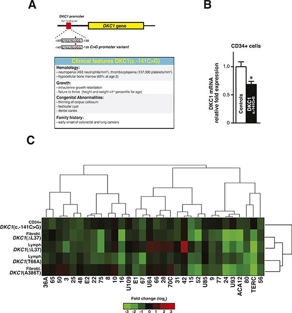
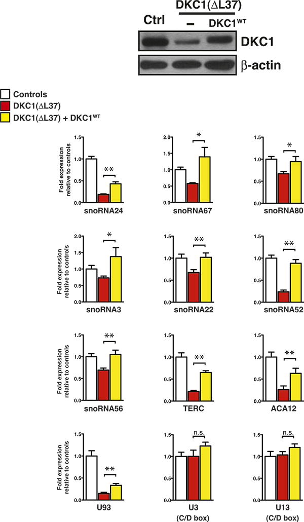
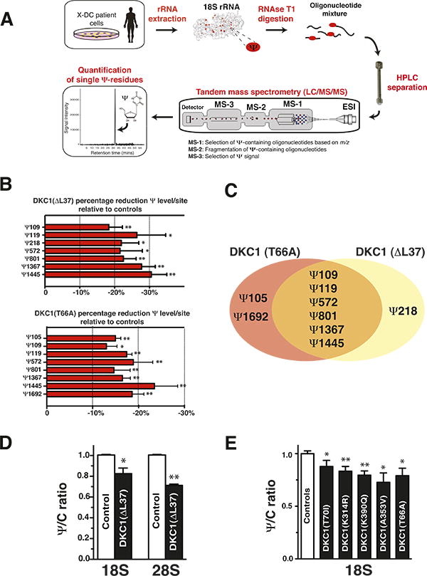
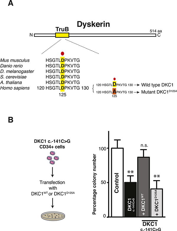
Noncoding RNAs control critical cellular processes, although their contribution to disease remains largely unexplored. Dyskerin associates with hundreds of H/ACA small RNAs to generate a multitude of functionally distinct ribonucleoproteins (RNPs). The DKC1 gene, encoding dyskerin, is mutated in the multisystem disorder X-linked dyskeratosis congenita (X-DC). A central question is whether DKC1 mutations affect the stability of H/ACA RNPs, including those modifying ribosomal RNA (rRNA). We carried out comprehensive profiling of dyskerin-associated H/ACA RNPs, revealing remarkable heterogeneity in the expression and function of subsets of H/ACA small RNAs in X-DC patient cells. Using a mass spectrometry approach, we uncovered single-nucleotide perturbations in dyskerin-guided rRNA modifications, providing functional readouts of small RNA dysfunction in X-DC. In addition, we identified that, strikingly, the catalytic activity of dyskerin is required for accurate hematopoietic stem cell differentiation. Altogether, these findings reveal that small noncoding RNA dysfunctions may contribute to the pleiotropic manifestation of human disease.


Figures

Figure 1
Figure 1. Characterization of dyskerin-associated H/ACA small RNAs in X-DC patient cells
(A) Schematic of the DKC1(c.-141C>G) mutation illustrating the position of the base pair substitution on the DKC1 promoter (top). Clinical features of the DKC1(c.-141C>G) patient are shown (bottom). (B) Quantification of DKC1 mRNA levels in CD34+ cells from two healthy controls and DKC1(c.-141C>G) patient was measured by real-time qPCR. (C) Heatmap diagram displaying hierarchical clustering of 32 H/ACA small RNA relative expression levels in X-DC patient cells. The expression of H/ACA small RNAs was measured by real-time qPCR, relative to two type-matched control cells for each mutation, and normalized to the abundance of RN7SK small non-coding RNA from at least three independent experiments. Each row represents a different type of X-DC patient-derived cell. Each column illustrates the expression of individual H/ACA small RNAs relative to controls. The color bar indicates the color-coding of small RNA expression from +3 to -3 in log2 space (bottom). Only slight variation in the expression of H/ACA small RNAs was observed amongst controls either by real-time qPCR or northern blot analysis (Figure S1 and Tables S1-S2). Statistical information is included in Table S2.
Figure 2
Figure 2. Expression of dyskerin rescues H/ACA small RNA expression levels in primary X-DC fibroblasts
Expression levels of different classes of small RNAs in control, DKC1(ΔL37) primary fibroblasts, and DKC1(ΔL37) primary fibroblasts expressing exogenous wild type dyskerin (DKC1WT) measured by real-time qPCR. Graphs show mean fold expression ± SEM relative to control and normalized to the abundance of RN7SK small non-coding RNA from three independent experiments. Western blot analysis of dyskerin expression is shown in control and DKC1(ΔL37) primary fibroblasts in the absence or presence of DKC1WT (top). β-actin was used as loading control. Statistical analysis was performed using the unpaired Student's t-test, *P<0.05 and **P<0.01.
Figure 3
Figure 3. Defective site-specific and global rRNA pseudouridylation manifest in X-DC patient cells
(A) Schematic representation of the mass spectrometry approach used to detect site-specific pseudouridine modifications on 18S rRNA from control and X-DC patient cells. m/z stands for mass-to-charge ratio. ESI is an abbreviation for electrospray ionization. (B) Site-specific quantification of Ψ levels in two controls and in DKC1(ΔL37) fibroblasts (top) and DKC1(T66A) lymphoblasts (bottom). Graph shows mean percentage Ψ reduction ± SEM relative to two controls at specific Ψ sites on 18S rRNA from three independent experiments. Quantification of all Ψ sites examined in this study is shown in Figures S3B-S3C. The accuracy of these measurements is well controlled for by performing calibration curves, demonstrating a linear response to different concentrations of synthetic Ψ-containing oligonucleotides within the range of Ψ values observed in patient samples (Figure S3D). The percentage reductions in Ψ measurements are relative and not absolute. (C) Venn diagram illustrates unique and commonly reduced Ψ sites in X-DC patient cells analyzed. (D) HPLC quantification of 18S and 28S rRNA Ψ levels in control and DKC1(ΔL37) primary fibroblasts. The graph shows mean Ψ to cytosine (Ψ/C) ratio ± SEM relative to controls from two independent experiments. (E) HPLC quantification of 18S rRNA Ψ levels in six independent controls and five X-DC lymphoblast cell lines harboring distinct DKC1 point mutations. The specific DKC1 mutation is presented on each column. The graph shows mean Ψ to cytosine (Ψ/C) ratio ± SEM for each X-DC lymphoblast cell line relative to controls from at least three independent experiments. Statistical analysis was performed using the unpaired Student's t-test, *P<0.05 and **P<0.01.
Figure 4
Figure 4. Dyskerin pseudouridylation activity is important for HSC differentiation
(A) Schematic presentation of dyskerin coding region highlighting evolutionary conservation of the TruB Ψ synthase domain across several species. A substitution of aspartic acid (D) with alanine (A) at position 125 abolishes dyskerin pseudouridylation activity and was employed as a catalytically inactive DKC1(D125A) mutant. (B) Hematopoietic colony forming assay was performed on CD34+ progenitor cells from a healthy control and DKC1(c.-141C>G) patient in the absence or presence of wild type DKC1WT or its catalytic mutant DKC1D125A using methylcellulose supplemented with complete cytokines cocktail sustaining the growth of all blood cell lineages. The total number of colonies formed was scored 10 days after plating. Graph shows the mean total percentage number of colonies ± SEM relative to controls from two independent experiments. Statistical analysis was performed using the unpaired Student's t-test, **P<0.01

References

    1. Alter BP, Baerlocher GM, Savage SA, Chanock SJ, Weksler BB, Willner JP, Peters JA, Giri N, Lansdorp PM. Very short telomere length by flow fluorescence in situ hybridization identifies patients with dyskeratosis congenita. Blood. 2007;110:1439–1447. - PMC - PubMed
    1. Alter BP, Giri N, Savage SA, Peters JA, Loud JT, Leathwood L, Carr AG, Greene MH, Rosenberg PS. Malignancies and survival patterns in the National Cancer Institute inherited bone marrow failure syndromes cohort study. British journal of haematology. 2010;150:179–188. - PMC - PubMed
    1. Apffel A, Chakel JA, Fischer S, Lichtenwalter K, Hancock WS. Analysis of Oligonucleotides by HPLC-Electrospray Ionization Mass Spectrometry. Analytical chemistry. 1997;69:1320–1325. - PubMed
    1. Bachellerie JP, Cavaille J. Guiding ribose methylation of rRNA. Trends in biochemical sciences. 1997;22:257–261. - PubMed
    1. Baerlocher GM, Vulto I, de Jong G, Lansdorp PM. Flow cytometry and FISH to measure the average length of telomeres (flow FISH) Nat Protoc. 2006;1:2365–2376. - PubMed

Publication types

MeSH terms