Review
. 2013 Jun;16(3):319-26.
doi: 10.1016/j.mib.2013.04.004. Epub 2013 May 23.

Inflammasome-mediated pyroptotic and apoptotic cell death, and defense against infection

Affiliations
Review

Inflammasome-mediated pyroptotic and apoptotic cell death, and defense against infection

Youssef Aachoui et al. Curr Opin Microbiol. 2013 Jun.

Abstract

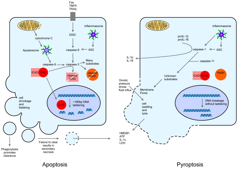
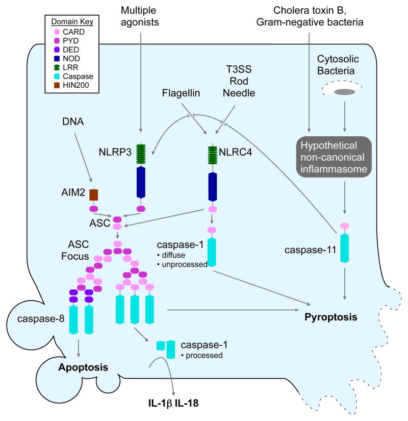
Cell death is an effective strategy to limit intracellular infections. Canonical inflammasomes, including NLRP3, NLRC4, and AIM2, recruit and activate caspase-1 in response to a range of microbial stimuli and endogenous danger signals. Caspase-1 then promotes the secretion of IL-1β and IL-18 and a rapid form of lytic programmed cell death termed pyroptosis. A second inflammatory caspase, mouse caspase-11, mediates pyroptotic death through an unknown non-canonical inflammasome system in response to cytosolic bacteria. In addition, recent work shows that inflammasomes can also recruit procaspase-8, initiating apoptosis. The induction of multiple pathways of cell death has probably evolved to counteract microbial evasion of cell death pathways.


Figures

Figure 1
Figure 1. The cellular events during pyroptosis and apoptosis
Both pyroptosis and apoptosis require activation of caspases, caspase-1/11 or caspase-2/3/7/8/9, respectively, but differ morphologically. Pyroptosis is characterized by formation of membrane pores between 1.1 and 2.4 nm in diameter. These pores likely cause dissipation of cellular ionic gradients leading to osmotic water influx, cell swelling, and plasma membrane lysis, releasing the cytosolic content into the extracellular space. Specific markers for this lysis are useful in vitro, for example lactate dehydrogenase release is readily detected by enzyme assay. Other cytosolic components released have inflammatory biologic functions, for example HMGB1, IL-1α, and ATP. On the other hand during apoptosis, plasma membrane integrity is maintained and cellular contents are not released. The plasma membrane loses leaflet lipid composition asymmetry as phosphatidyl serine becomes exposed on the outer leaflet, promoting phagocytosis. However, in the absence of phagocytosis, at later timepoints the apoptotic bodies undergo secondary necrosis characterized by rupture of the membrane. Apoptosis is an energy requiring process during which cells shrink and form apoptotic blebs. The DNA repair factor PARP1 is inactivated by cleavage, and nuclear condensation and fragmentation occur. Internucleosomal DNA cleavage, detected as laddering on gel electrophoresis or by TUNEL staining for DNA ends, occurs due to ICAD degradation and activation of CAD (caspase-activated DNase). Weak TUNEL staining is also seen in pyroptosis, but DNA laddering and ICAD degradation are absent [55].
Figure 2
Figure 2. Canonical and non-canonical inflammasome pathways to cell death and cytokine secretion
Canonical inflammasomes are cytosolic platforms that activate caspase-1 in response to pathogen- or danger- associated molecular patterns. Activation of caspase-1 leads to secretion of IL-1β and IL-18, and pyroptosis. Two domain structures for inflammasome initiator proteins are known. 1) NLRs contain a signaling domain (PYD or CARD), a nucleotide binding oligomerization domain (NOD/NACHT), and a leucine rich repeat (LRR) domain. 2) AIM2 contains a PYD signaling domain and a DNA-binding HIN-200 domain. AIM2 detects cytosolic DNA. NLRP3 detects multiple agonists such as extracellular ATP, bacterial pore-forming toxins, monosodium urate crystals, and cholesterol crystals. NLRC4 detects cytosolic flagellin or T3SS components that have been injected into the cytosol. The NLR or AIM2 signaling domains (PYD or CARD) bind to ASC by homotypic interactions, triggering formation of the ASC focus, which recruits procaspase-1, leading to its activation and processing. The ASC focus also recruits procaspase-8, initiating an apoptotic pathway. NLRC4 can additionally interact directly with caspase-1, resulting only in pyroptosis. The platform(s) that activate caspase-11 in response to cytosolic bacteria remain unknown. Caspase-11 triggers pyroptosis directly, and can also activate the canonical NLRP3-ASC-caspase-1 inflammasome pathway leading to IL-1β and IL-18 processing (denoted by the long curved arrow to NLRP3).

References

    1. Miao EA, Rajan JV, Aderem A. Caspase-1-induced pyroptotic cell death. Immunol Rev. 2011;243:206–214. - PMC - PubMed
    1. Moltke Von J, Ayres JS, Kofoed EM, Chavarría-Smith J, Vance RE. Recognition of Bacteria by Inflammasomes. Annu Rev Immunol. 2012 doi: 10.1146/annurev-immunol-032712-095944. - DOI - PubMed
    1. Angosto D, López-Castejón G, López-Muñoz A, Sepulcre MP, Arizcun M, Meseguer J, Mulero V. Evolution of inflammasome functions in vertebrates: Inflammasome and caspase-1 trigger fish macrophage cell death but are dispensable for the processing of IL-1β. Innate Immunity. 2012;18:815–824. - PubMed
    1. Ashida H, Mimuro H, Ogawa M, Kobayashi T, Sanada T, Kim M, Sasakawa C. Cell death and infection: a double-edged sword for host and pathogen survival. The Journal of Cell Biology. 2011;195:931–942. - PMC - PubMed
    1. Vanlangenakker N, Vanden Berghe T, Vandenabeele P. Many stimuli pull the necrotic trigger, an overview. Cell Death Differ. 2011;19:75–86. - PMC - PubMed

Publication types