

Pushing the limits of in vivo diffusion MRI for the Human Connectome Project
- PMID: 23707579
- PMCID: PMC3725309
- DOI: 10.1016/j.neuroimage.2013.05.078
Pushing the limits of in vivo diffusion MRI for the Human Connectome Project
Abstract
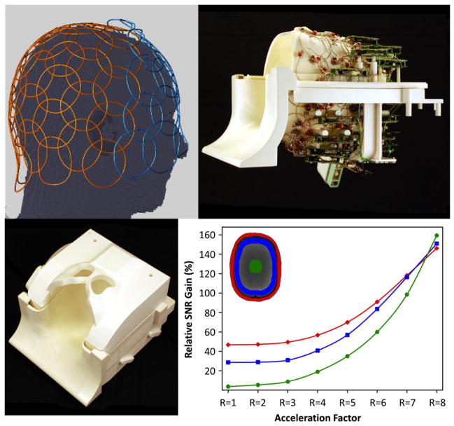
Perhaps more than any other "-omics" endeavor, the accuracy and level of detail obtained from mapping the major connection pathways in the living human brain with diffusion MRI depend on the capabilities of the imaging technology used. The current tools are remarkable; allowing the formation of an "image" of the water diffusion probability distribution in regions of complex crossing fibers at each of half a million voxels in the brain. Nonetheless our ability to map the connection pathways is limited by the image sensitivity and resolution, and also the contrast and resolution in encoding of the diffusion probability distribution. The goal of our Human Connectome Project (HCP) is to address these limiting factors by re-engineering the scanner from the ground up to optimize the high b-value, high angular resolution diffusion imaging needed for sensitive and accurate mapping of the brain's structural connections. Our efforts were directed based on the relative contributions of each scanner component. The gradient subsection was a major focus since gradient amplitude is central to determining the diffusion contrast, the amount of T2 signal loss, and the blurring of the water PDF over the course of the diffusion time. By implementing a novel 4-port drive geometry and optimizing size and linearity for the brain, we demonstrate a whole-body sized scanner with G(max) = 300 mT/m on each axis capable of the sustained duty cycle needed for diffusion imaging. The system is capable of slewing the gradient at a rate of 200 T/m/s as needed for the EPI image encoding. In order to enhance the efficiency of the diffusion sequence we implemented a FOV shifting approach to Simultaneous MultiSlice (SMS) EPI capable of unaliasing 3 slices excited simultaneously with a modest g-factor penalty allowing us to diffusion encode whole brain volumes with low TR and TE. Finally we combine the multi-slice approach with a compressive sampling reconstruction to sufficiently undersample q-space to achieve a DSI scan in less than 5 min. To augment this accelerated imaging approach we developed a 64-channel, tight-fitting brain array coil and show its performance benefit compared to a commercial 32-channel coil at all locations in the brain for these accelerated acquisitions. The technical challenges of developing the over-all system are discussed as well as results from SNR comparisons, ODF metrics and fiber tracking comparisons. The ultra-high gradients yielded substantial and immediate gains in the sensitivity through reduction of TE and improved signal detection and increased efficiency of the DSI or HARDI acquisition, accuracy and resolution of diffusion tractography, as defined by identification of known structure and fiber crossing.
Keywords: DSI; Diffusion imaging; Gradient hardware; HARDI; MRI; Structural connectivity.
Published by Elsevier Inc.
Figures


References
-
- Guidelines for limiting exposure to time-varying electric and magnetic fields (1 Hz to 100 kHz) Health physics. 99(6):818–836. - PubMed
-
- Aganj I, Lenglet C, Sapiro G, Yacoub E, Ugurbil K, Harel N. Multiple Q-shell ODF reconstruction in Q-ball imaging. Medical image computing and computer-assisted intervention: MICCAI … International Conference on Medical Image Computing and Computer-Assisted Intervention. 2009;12(Pt 2):423–431. - PMC - PubMed
-
- Alagappan V, Nistler J, Adalsteinsson E, Setsompop K, Fontius U, Zelinski A, Vester M, Wiggins GC, Hebrank F, Renz W, Schmitt F, Wald LL. Degenerate mode band-pass birdcage coil for accelerated parallel excitation. Magn Reson Med. 2007;57(6):1148–1158. - PubMed
-
- Alford JK, Rutt BK, Scholl TJ, Handler WB, Chronik BA. Delta relaxation enhanced MR: improving activation-specificity of molecular probes through R1 dispersion imaging. Magnetic resonance in medicine: official journal of the Society of Magnetic Resonance in Medicine/Society of Magnetic Resonance in Medicine. 2009;61(4):796–802. - PubMed
-
- Anderson AW. Measurement of fiber orientation distributions using high angular resolution diffusion imaging. Magn Reson Med. 2005;54(5):1194–1206. - PubMed
Publication types
MeSH terms
Grants and funding
LinkOut - more resources
Full Text Sources
Other Literature Sources
Medical
Miscellaneous
