Review
. 2013 May 24;13(6):6882-99.
doi: 10.3390/s130606882.

Advanced respiratory motion compensation for coronary MR angiography

Affiliations
Review

Advanced respiratory motion compensation for coronary MR angiography

Markus Henningsson et al. Sensors (Basel). .

Abstract

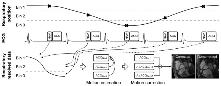
Despite technical advances, respiratory motion remains a major impediment in a substantial amount of patients undergoing coronary magnetic resonance angiography (CMRA). Traditionally, respiratory motion compensation has been performed with a one-dimensional respiratory navigator positioned on the right hemi-diaphragm, using a motion model to estimate and correct for the bulk respiratory motion of the heart. Recent technical advancements has allowed for direct respiratory motion estimation of the heart, with improved motion compensation performance. Some of these new methods, particularly using image-based navigators or respiratory binning, allow for more advanced motion correction which enables CMRA data acquisition throughout most or all of the respiratory cycle, thereby significantly reducing scan time. This review describes the three components typically involved in most motion compensation strategies for CMRA, including respiratory motion estimation, gating and correction, and how these processes can be utilized to perform advanced respiratory motion compensation.


Figures

Figure 1.
Figure 1.
CMRA dataset without respiratory motion correction (a) and with respiratory motion correction (b). The 3D dataset is reformatted to visualize the right coronary artery (RCA) and left anterior descending artery (LAD). Ao = ascending aorta; RV = right ventricle; LV = left ventricle. Adapted from [11].
Figure 2.
Figure 2.
Schematics of ECG-triggered CMRA sequence. One k-space data segment is acquired (ACQ) following a time delay (trigger delay) after the R-wave to minimize cardiac motion artifacts. Typically the trigger delay is adjusted to coincide with the mid-diastolic rest period and the center of k-space. Preparation pulses (PP) can be used to improve visualization of the coronary arteries by suppressing signal from surrounding tissues, including epicardial fat and myocardium. Respiratory motion compensation (RMC), including motion estimation, gating and correction, is typically performed prior to ACQ, although gating and correction may be also being performed retrospectively.
Figure 3.
Figure 3.
Scan planning of d1D NAV in axial plane using either a 2D selective RF “pencil beam” excitation (a) or a spin-echo approach with obliquely aligned 90° excitation and 180° refocusing pulses (b). The d1D NAV positioned in the coronal plane, on the dome of the right hemi-diaphragm with the readout in foot-head (FH) direction (c). The d1D NAV signal (d) clearly captures the displacement of the lung-liver interface along the FH direction (dFH) over time (t).
Figure 4.
Figure 4.
Sequence diagram of two-dimensional image-based self-navigation (2D NAV) using the startup echoes of a 3D balanced Steady-State Free Precession (bSSFP) sequence (a). The 2D NAV is generated by adding phase encoding gradients (Gphase) to the startup echoes, using a high-low profile order. The number of startup echoes is proportional to the 2D NAV phase encoding resolution, however 10 startup echoes are commonly used which results in accurate respiratory motion estimation of the heart. Whole-heart CMRA from two healthy volunteers using either the conventional diaphragmatic 1D navigator (d1D NAV) with a tracking factor of 0.6 for motion estimation (top row) or 2D NAV (bottom row) with improved coronary vessel sharpness of the distal LAD (arrows) for the 2D NAV approach (b). Adapted from [65].
Figure 5.
Figure 5.
Respiratory 1D navigator signals throughout a scan. One column represents one navigator acquisition, and the red dot the measured respiratory position. Image data acquired at positions outside of the gating window, shown by the blue lines, are rejected and re-acquired in the following shot and only accepted once the corresponding navigator has been acquired within the gating window which is signified by the green lines at the bottom of each navigator image. Although this method effectively reduces the respiratory motion artifacts, it prolongs the scan, typically by a factor of 2 or more.
Figure 6.
Figure 6.
Schematic of a general respiratory binning, motion estimation and correction procedure. The respiratory position is measured using a navigator (NAV) for every data acquisition (ACQ), which can then be associated to a respiratory bin. As respiration is cyclical this allows multiple ACQs to be associated to a particular respiratory bin. Data from each bin can be reconstructed separately (ACQBin1-3) and is now respiratory resolved, although undersampled. For radial binning approaches the undersampled data for each bin can be used to estimate motion between bins [67,68]. However, for Cartesian binning approaches additional fully sampled data can be acquired [11,69]. The respiratory motion is typically estimated by registering ACQBin2 and ACQBin3 to the end-expiratory ACQBin1 to generate the transformations A2 and A3. The motion corrected CMRA data can be obtained by applying these transformations to the CMRA data in the corresponding bins and summing the results. Note, in this example 3 respiratory bins are used, however in practice 4 to 6 bins are typically employed. Adapted from [67].

References

    1. Go A.S., Mozaffarian D., Roger V.L., Benjamin E.J., Berry J.D., Borden W.B., Bravata D.M., Dai S., Ford E.S., Fox C.S., et al. Heart disease and stroke statistics—2013 update: A report from the american heart association. Circulation. 2013;127:e6–e245. - PMC - PubMed
    1. Dewey M. Coronary CT versus MR angiography: Pro CT—The role of CT angiography. Radiology. 2011;258:329–339. - PubMed
    1. Wang Y., Riederer S.J., Ehman R.L. Respiratory motion of the heart-kinematics and the implications for the spatial-resolution in coronary imaging. Magn. Reson. Med. 1995;33:713–719. - PubMed
    1. Keegan J., Gatehouse P., Yang G.Z., Firmin D. Coronary artery motion with the respiratory cycle during breath-holding and free-breathing: Implications for slice-followed coronary artery imaging. Magn. Reson. Med. 2002;47:476–481. - PubMed
    1. Danias P.G., Stuber M., Botnar R.M., Kissinger K.V., Edelman R.R., Manning W.J. Relationship between motion of coronary arteries and diaphragm during free breathing: Lessons from real-time MR imaging. Am. J. Roentgenol. 1999;172:1061–1065. - PubMed

MeSH terms