. 2013 Aug 7;3(8):1261-72.
doi: 10.1534/g3.113.006213.

Recombination-induced tag exchange (RITE) cassette series to monitor protein dynamics in Saccharomyces cerevisiae

Affiliations

Recombination-induced tag exchange (RITE) cassette series to monitor protein dynamics in Saccharomyces cerevisiae

Marit Terweij et al. G3 (Bethesda). .

Abstract

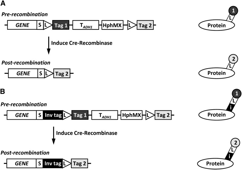
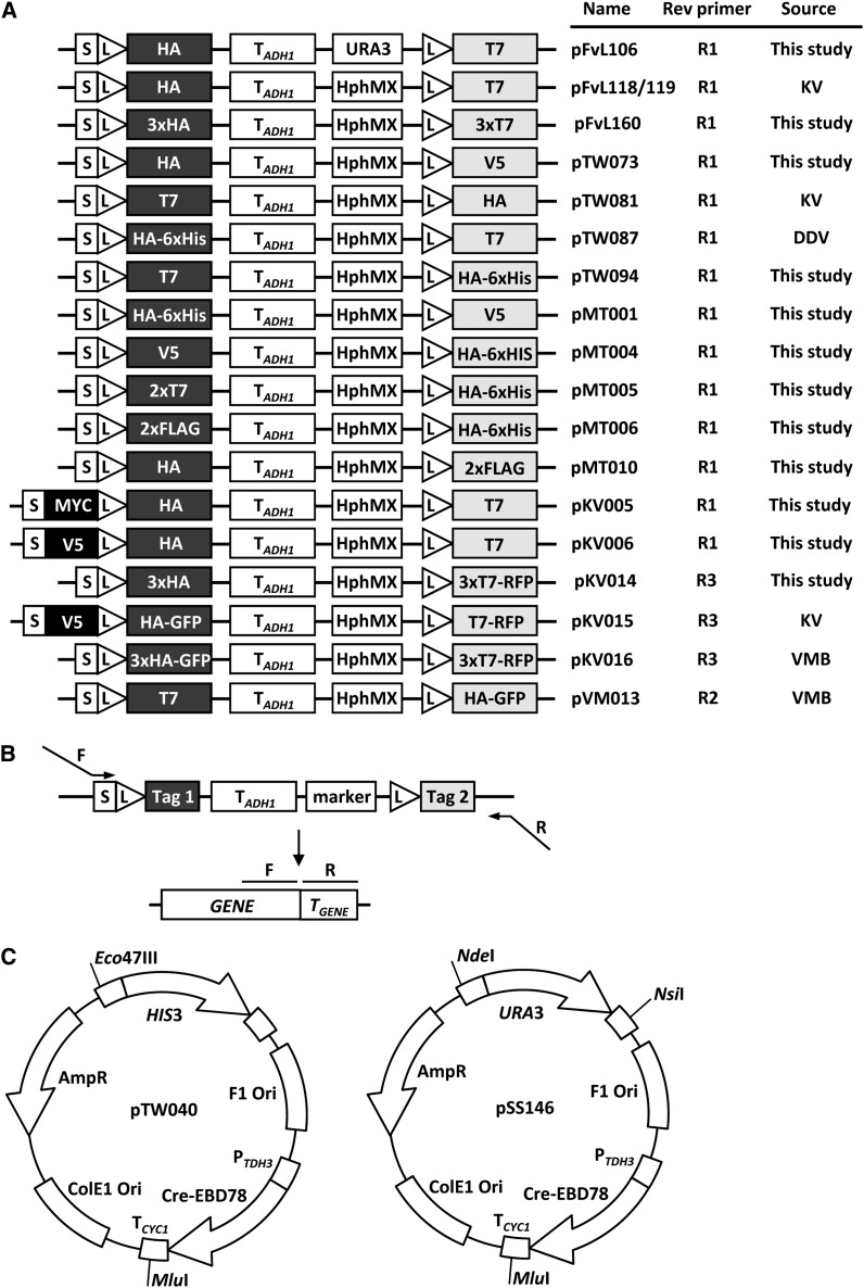
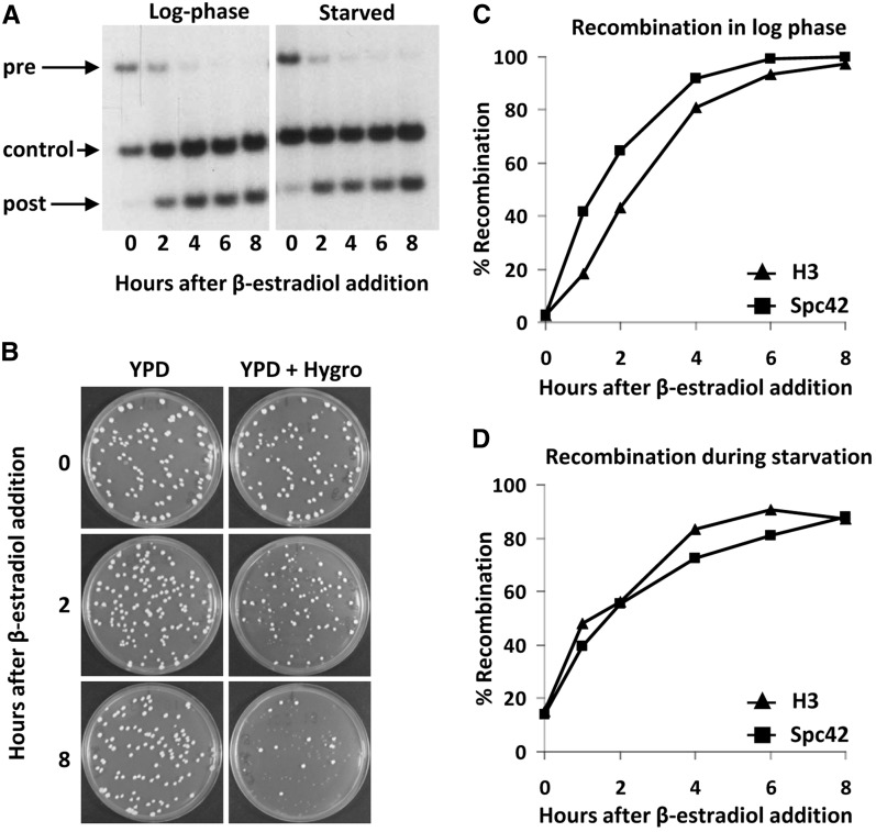
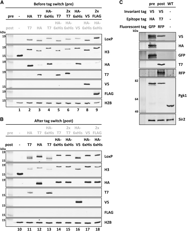
Proteins are not static entities. They are highly mobile, and their steady-state levels are achieved by a balance between ongoing synthesis and degradation. The dynamic properties of a protein can have important consequences for its function. For example, when a protein is degraded and replaced by a newly synthesized one, posttranslational modifications are lost and need to be reincorporated in the new molecules. Protein stability and mobility are also relevant for the duplication of macromolecular structures or organelles, which involves coordination of protein inheritance with the synthesis and assembly of newly synthesized proteins. To measure protein dynamics, we recently developed a genetic pulse-chase assay called recombination-induced tag exchange (RITE). RITE has been successfully used in Saccharomyces cerevisiae to measure turnover and inheritance of histone proteins, to study changes in posttranslational modifications on aging proteins, and to visualize the spatiotemporal inheritance of protein complexes and organelles in dividing cells. Here we describe a series of successful RITE cassettes that are designed for biochemical analyses, genomics studies, as well as single cell fluorescence applications. Importantly, the genetic nature and the stability of the tag switch offer the unique possibility to combine RITE with high-throughput screening for protein dynamics mutants and mechanisms. The RITE cassettes are widely applicable, modular by design, and can therefore be easily adapted for use in other cell types or organisms.

Keywords: epitope tag; protein inheritance; protein turnover; pulse-chase.


Figures

Figure 1
Figure 1
Outline of RITE. (A) After integration of a RITE cassette behind the gene of interest (GENE), recombination between LoxP sites is induced by Cre-Recombinase, causing a permanent switch from old Tag 1 to new Tag 2 on the protein of interest. S, spacer; L, LoxP recombination sites; TADH1, ADH1 terminator; HphMX, Hygromycin resistance cassette. (B) RITE cassette including an invariant tag (i) upstream of the first LoxP site. The invariant tag is present pre- and postrecombination and can be used for simultaneous detection of the old and new protein of interest.
Figure 2
Figure 2
RITE cassettes and Cre recombinase vectors. (A) Available RITE cassettes. The cassettes can be amplified with the primers indicated (see panel B). The sequence of the forward primer F1 is 5′-GGT GGA TCT GGT GGA TCT-3′ (corresponding to the reading frame of the spacer sequence); the sequences of the reverse primers are: R1 5′-AGGGAACAAAAGCTTGCATG-3′; R2 5′-TGATTACGCCAAGCTCG-3′; R3 5′-TCA GGCGCCGGTGGAGTGGCG-3′. Note that primer R3 contains a STOP codon (underlined), because the mRFP sequence in pKV14-16 lacks a stop codon. (B) Gene targeting of RITE cassettes by homologous recombination. The RITE cassettes can be PCR-amplified with a forward and reverse primer that have 20 bp homology with the cassette and a tail of at least 40 bp homology with the 3′ end of the gene of interest and 3′ UTR, respectively. (C) Constructs to integrate the Cre recombinase expression vector in the yeast genome by homologous recombination. Unique restriction sites can be used to digest the plasmid and integrate the linear fragment by single cross-over (see Materials and Methods).
Figure 3
Figure 3
Cre recombination kinetics. (A) Southern blot analysis of H3-HA→T7 (strain NKI2048). Recombination was induced in log-phase or nutrient-starved cells and analyzed using an H3-specific probe on HindIII-digested DNA. The three bands recognized are specific for preswitch (pre), postswitch (post), or an internal control present in both (control). (B) Plating assay of starved Spc42-3xHA→3xT7-RFP cells (strain SPC42R) after induction of Cre-recombination. Cells were plated on YEPD (YPD) media and then replica plated to YEPD media containing Hygromycin (Hygro). Quantification of recombination levels as measured by plating assay of H3-V5→HA-6xHIS (strain NKI8050) and strain SPC42R in log phase (C) and during starvation (D).
Figure 4
Figure 4
Immunoblot analysis of RITE-tagged histone H3 and Pgk1. (A) Histone H3 (HHT2) was tagged with different RITE cassettes. Before tag switch (pre) detects the old Tag 1. A wild-type strain (−, untagged H3) was used as a negative control. H2B was used as a loading control. (B) After tag switch (post) detects the new Tag 2. Postswitch strains are the recombined counterparts of the preswitch strains shown in (A). Strains used: 1. NKI8001; 2. NKI2148; 3. NKI2158; 4. NKI2178; 5. NKI2220; 6. NKI8051; 7. NKI8056; 8. NKI8050; 9. NKI8052; 10. NKI8001; 11. NKI8085; 12. NKI4138; 13. NKI8086; 14. NKI8037; 15. NKI8089; 16. NKI8058; 17. NKI8087; 18. NKI8088. (C) Pgk1 was tagged with an HA-yEGFP→T7-mRFP RITE cassette containing an invariant epitope tag V5 (V5i). Sir2 was used as loading control.
Figure 5
Figure 5
Validation of RITE tags in ChIP assays. RITE-ChIP of H3 and H2A.Z followed by qPCR analysis of three loci: A = PTC6, B = RSA4, C = SPA2. The regions around the transcription start sites of PTC6 and RSA4 have high H2A.Z occupancy; the SPA2 region is located at the 5′ end of the SPA2 coding sequence and has low H2A.Z occupancy (primer sequences are listed in Table 3). ChIP signals were normalized over the corresponding inputs. The relative ChIP efficiency (H2A.Z/H3 on each of the three loci examined) varied between the four epitope tags. Average of three biological replicates ± SEM. Results were obtained from: HA-ChIP on strains NKI2178 (H3-HA-6xHIS→T7) and NKI8030 (H2A.Z-HA-6xHIS→T7), V5-ChIP on strains NKI8050 (H3-V5→HA-6xHIS) and NKI8053 (H2A.Z-V5→HA-6xHIS), FLAG-ChIP on strains NKI8052 (H3-2xFLAG→HA-6xHIS) and NKI8055 (H2A.Z-2xFLAG→HA-6xHIS), and T7-ChIP on strains NKI8051 (H3-2xT7→HA-6xHIS) and NKI8054 (H2A.Z-2xT7→HA-6xHIS.
Figure 6
Figure 6
Analysis of fluorescent knock-in RITE cassettes. (A) Spc42, a subunit of the spindle pole body, was tagged with a 3xHA→3xT7-mRFP RITE cassette (strain Spc42R). (B) Fluorescent microscopy of strain Spc42R. In the preswitch sample, no mRFP was detected; in the postswitch sample Spc42-mRFP was clearly visible. (C) Immunoblot analysis of the Spc42R samples described in (B). Pgk1 was used as loading control. (D−F) as in (A−C) but using Spc42 tagged with a 1xT7→1xHA-yEGFP RITE cassette (strain Spc42G). Spc42 containing a single HA or T7 tag could not be detected on blots using the tag-specific antibodies. However, an antibody against spacer-LoxP could visualize old and new Spc42. Scale bars, 2 μm.
Figure 7
Figure 7
Immunoblot and ChIP analysis of histone H3 dynamics. (A) Histone H3 was tagged with a V5→HA-6xHis RITE cassette in strain NKI8050. (B) Outline of experimental setup. (C) Immunoblot analysis of cells arrested by starvation (pre), switched for 16 hr (post), and subsequently released into fresh media for 2 hr (no cell division) or 4 hr (one cell division). An antibody against spacer-LoxP detects both old and new H3. (D-E) H3-V5 and H3-HA-6xHis ChIP analysis of samples described in (B) to measure occupancy (IP/input) of old and new H3 at PTC6 (around transcription start site) and SPA2 (coding sequence) (primer sequences are listed in Table 3). (F) Exchange of histone H3 determined as the ChIP ratio of new H3 over old H3 (HA/V5). Average of three biological replicates ± SEM.

References

    1. Adams S. R., Tsien R. Y., 2008. Preparation of the membrane-permanent biarsenicals FlAsH-EDT2 and ReAsH-EDT2 for fluorescent labeling of tetracysteine-tagged proteins. Nat. Protoc. 3: 1527–1534 - PMC - PubMed
    1. Albert I., Mavrich T. N., Tomsho L. P., Qi J., Zanton S. J., et al. , 2007. Translational and rotational settings of H2A.Z nucleosomes across the Saccharomyces cerevisiae genome. Nature 446: 572–576 - PubMed
    1. Brachmann C. B., Davies A., Cost G. J., Caputo E., Li J., et al. , 1998. Designer deletion strains derived from Saccharomyces cerevisiae S288C: a useful set of strains and plasmids for PCR-mediated gene disruption and other applications. Yeast 14: 115–132 - PubMed
    1. Butko M. T., Yang J., Geng Y., Kim H. J., Jeon N. L., et al. , 2012. Fluorescent and photo-oxidizing TimeSTAMP tags track protein fates in light and electron microscopy. Nat. Neurosci. 15: 1742–1751 - PMC - PubMed
    1. Deal R., Henikoff S., 2010a Capturing the dynamic epigenome. Genome Biol. 11: 218. - PMC - PubMed

Publication types

MeSH terms