. 2013 Aug;34(8):1101-7.
doi: 10.1038/aps.2013.48. Epub 2013 May 27.

Dihydroartemisinin promotes angiogenesis during the early embryonic development of zebrafish

Affiliations

Dihydroartemisinin promotes angiogenesis during the early embryonic development of zebrafish

Qian Ba et al. Acta Pharmacol Sin. 2013 Aug.

Abstract

Aim: To investigate the embryotoxicity of dihydroartemisinin (DHA), the main active metabolite of artemisinin, in zebrafish, and explore the corresponding mechanisms.

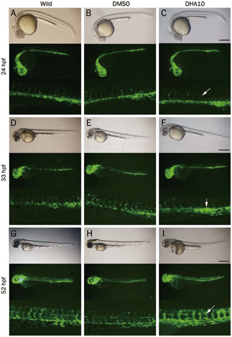
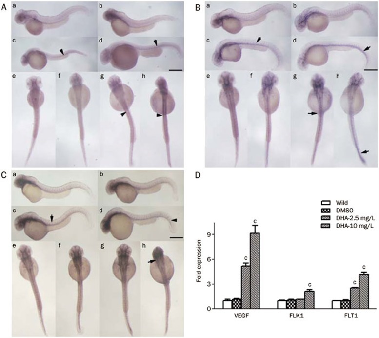
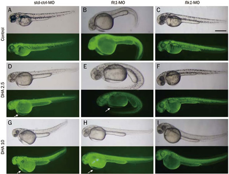
Methods: The embryos of wild type and TG (flk1:GFP) transgenic zebrafish were exposed to DHA. Developmental phenotypes of the embryos were observed. Development of blood vessels was directly observed in living embryos of TG (flk1:GFP) transgenic zebrafish under fluorescence microscope. The expression of angiogenesis marker genes vegfa, flk1, and flt1 in the embryos was detected using real-time PCR and RNA in situ hybridization assays.

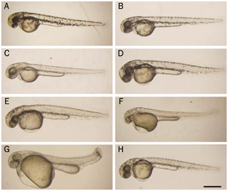
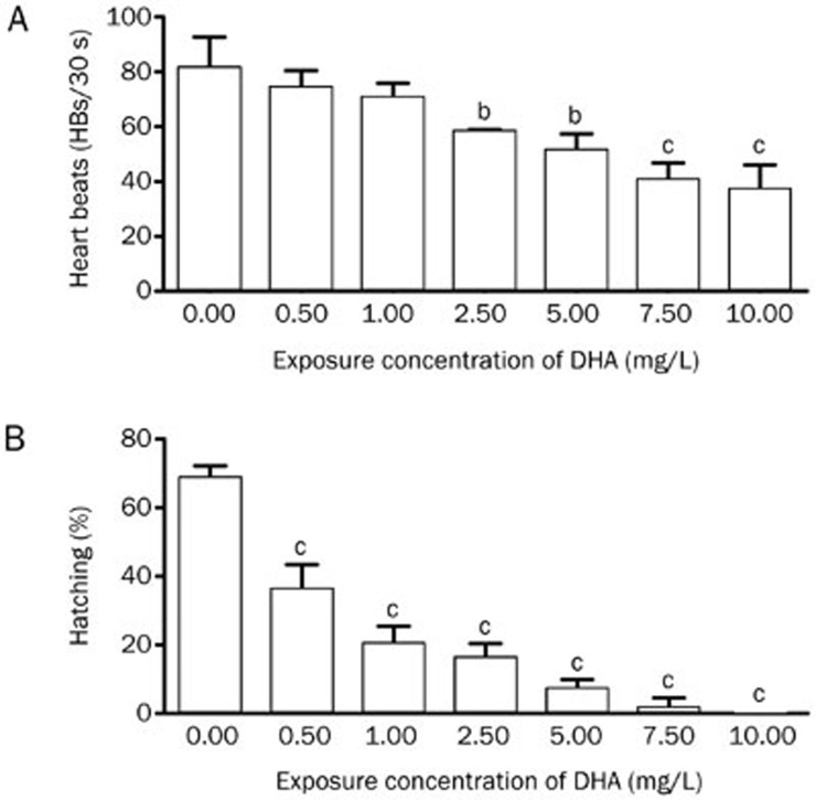
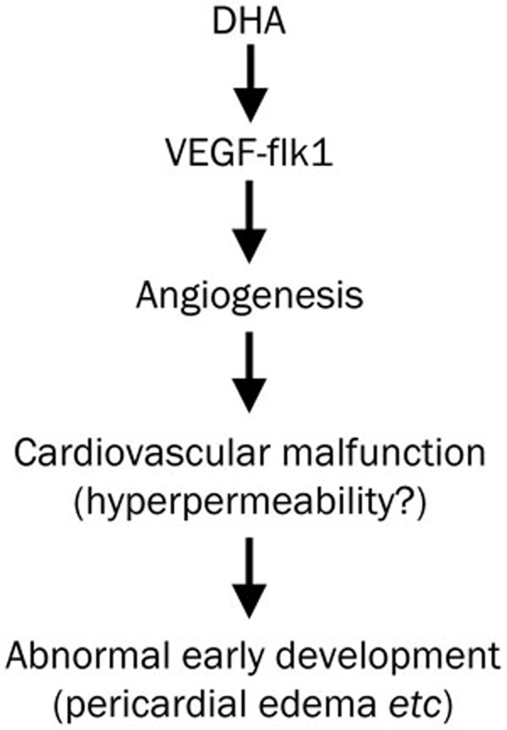
Results: Exposure to DHA (1-10 mg/L) dose-dependently caused abnormal zebrafish embryonic phenotypes in the early developmental stage. Furthermore, exposure to DHA (10 mg/L) resulted in more pronounced embryonic angiogenesis in TG (flk1:GFP) zebrafish line. Exposure to DHA (10 mg/L) significantly increased the mRNA expression of vegfa, flk1, and flt1 in the embryos. Knockdown of the flk1 protein partially blocked the effects of DHA on embryogenesis.

Conclusion: DHA causes abnormal embryonic phenotypes and promotes angiogenesis in zebrafish early embryonic development, demonstrating the potential embryotoxicity of DHA.


Figures

Figure 1
Figure 1
Morphological changes induced by dihydroartemisinin (DHA) and artemisinin during zebrafish early embryonic development. Embryos treated with different concentrations of compounds were imaged at 48 hpf. (A) No treatment; (B) 0.1% DMSO; (C) 1.0 mg/L DHA; (D) 2.5 mg/L DHA; (E) 5.0 mg/L DHA; (F) 10 mg/L DHA; (G) 15 mg/L DHA; (H) 10 mg/L artemisinin. The scale bar represents 500 μm for all panels.
Figure 2
Figure 2
The heartbeats and hatching success of embryos exhibit a dose-dependent decrease. (A) Embryos were treated, and the heartbeats were examined at 60 hpf. Three replicates of 60 embryos were used for each concentration group. (B) A total of 60 embryos were used per group. Shown are results representative from three independent experiments. bP<0.05, cP<0.01 compared with the control embryos. DHA, dihydroartemisinin.
Figure 3
Figure 3
Embryos from the TG (flk1:GFP) zebrafish line display angiogenesis abnormalities with dihydroartemisinin (DHA) exposure. Three groups of embryos were used to compare changes including the untreated, DMSO-treated and DHA-treated (10 mg/L DHA) groups, and different developmental stages were selected to show changes, including 24 hpf (A, B, C), 33 hpf (D, E, F), and 55 hpf (G, H, I). The white arrows in panels C, F, and I indicate abnormalities in embryos compared with untreated and DMSO-treated embryos. The embryos shown in all panels are lateral views. The scale bar represents 500 μm for the first and second row in each panel and 1500 μm for the third row in each panel.
Figure 4
Figure 4
Dihydroartemisinin (DHA) enhances the expression of angiogenesis marker genes in zebrafish embryonic development. (A–C) The WISH results for vegfa (A), flk1 (B), and flt1 (C) expression induced by DHA during early embryogenesis at 48 hpf. The embryos in panels A (a, e), B (a, e), and C (a, e) were untreated, the embryos in panels A (b, f), B (b, f), and C (b, f) were DMSO-treated, the embryos in panels A (c, g), B (c, g), and C (c, g) were treated with 2.5 mg/L DHA, and the embryos in panels A (d, h), B (d, h), and C (d, h) were treated with 10 mg/L DHA. Panels A (a–d), B (a–d), C (a–d) are lateral views and the others are dorsal views. The scale bar represents 650 μm for panels A (a–d), B (a–d), C (a–d), and 150 μm for panels A (e–h), B (e–h), C (e–h). (D) The mRNA expression level of vegfa, flk1, and flt1 at 48 hpf was examined by real-time PCR. Data shown are the mean±SEM for at least three independent experiments. cP<0.01 compared with the control embryos.
Figure 5
Figure 5
Antisense knockdown of flk1 in zebrafish abolishes dihydroartemisinin (DHA)-induced pericardial edema. (A–C) Embryos were treated with DMSO. (D–F) Embryos were treated with 2.5 mg/L DHA. (G–I) Embryos were treated with 10 mg/L DHA. NC-MO injected embryos are shown in panels A, D, and G. flt1-MO injected embryos are shown in panels B, E, and H. flk1-MO injected embryos are shown in panels C, F, and I. The scale bar represents 500 μm for all panels.
Figure 6
Figure 6
The proposed mechanism by which dihydroartemisinin (DHA) exposure causes abnormal early embryonic development in zebrafish.

References

    1. Cui L, Su XZ. Discovery, mechanisms of action and combination therapy of artemisinin. Expert Rev Anti Infect Ther. 2009;7:999–1013. - PMC - PubMed
    1. Klayman DL. Qinghaosu (artemisinin): an antimalarial drug from China. Science. 1985;228:1049–55. - PubMed
    1. Chen T, Li M, Zhang R, Wang H. Dihydroartemisinin induces apoptosis and sensitizes human ovarian cancer cells to carboplatin therapy. J Cell Mol Med. 2009;13:1358–70. - PMC - PubMed
    1. Firestone GL, Sundar SN. Anticancer activities of artemisinin and its bioactive derivatives. Expert Rev Mol Med. 2009;11:e32. - PubMed
    1. Nakase I, Lai H, Singh NP, Sasaki T. Anticancer properties of artemisinin derivatives and their targeted delivery by transferrin conjugation. Int J Pharm. 2008;354:28–33. - PubMed

Publication types

MeSH terms

LinkOut - more resources