Comparative Study
. 2013 Jul;20(7):827-35.
doi: 10.1038/nsmb.2593. Epub 2013 May 26.

Electron microscopy structure of human APC/C(CDH1)-EMI1 reveals multimodal mechanism of E3 ligase shutdown

Affiliations
Comparative Study

Electron microscopy structure of human APC/C(CDH1)-EMI1 reveals multimodal mechanism of E3 ligase shutdown

Jeremiah J Frye et al. Nat Struct Mol Biol. 2013 Jul.

Abstract

The anaphase-promoting complex/cyclosome (APC/C) is a ~1.5-MDa multiprotein E3 ligase enzyme that regulates cell division by promoting timely ubiquitin-mediated proteolysis of key cell-cycle regulatory proteins. Inhibition of human APC/C(CDH1) during interphase by early mitotic inhibitor 1 (EMI1) is essential for accurate coordination of DNA synthesis and mitosis. Here, we report a hybrid structural approach involving NMR, electron microscopy and enzymology, which reveal that EMI1's 143-residue C-terminal domain inhibits multiple APC/C(CDH1) functions. The intrinsically disordered D-box, linker and tail elements, together with a structured zinc-binding domain, bind distinct regions of APC/C(CDH1) to synergistically both block the substrate-binding site and inhibit ubiquitin-chain elongation. The functional importance of intrinsic structural disorder is explained by enabling a small inhibitory domain to bind multiple sites to shut down various functions of a 'molecular machine' nearly 100 times its size.


Figures
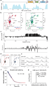
Figure 1
Figure 1. The APC/C inhibitory domain of EMI1 contains two intrinsically disordered segments separated by a zinc-dependent folded domain
a, Output of structure prediction program PONDR, with schematic views of EMI1 structural elements shown above. F - F-box–SKP1, D - D-box, L - Linker, Z - Zinc Binding Region (ZBR), T- Tail. b, Overlay of 15N-1H HSQC spectra of uniformly 15N-labeled EMI1DLZT (blue, consisting of the D-box, Linker, Zinc Binding Region (ZBR), and Tail, corresponding to the APC/CCDH1 inhibitory domain12,22), with EMI1ZT (red) or EMI1Z (green),superimposition showing dispersal of peaks found in the isolated EMI1Z domain. A subset of disperse resonances assigned for EMI1DLZT are labeled. c, Heteronuclear NOE data and difference in Cα chemical shifts (measured – predicted for random coil 50) for uniformly 15N-labeled EMI1DLZT measured at 25°C. Similar results obtained at 5°C are not shown. d, Overlay of 15N-1H HSQC spectrum of EMI1ZT (red) with that after treatment with EDTA (black) on left, and after subsequent reconstitution with zinc (blue) on right. e, 1D proton NMR spectra for EMI1ZT or the indicated cysteine mutants. f, Dynamic light scattering data (DynaPro Nanostar) and fit for hydrodynamic radius (Dynamics V7.1.7 software, Wyatt) for EMI1DLZT. g, Analytical ultracentrifugation data showing sedimentation coefficient distribution c(s) analysis for determination of sedimentation coefficient of EMI1DLZT, for calculation of hydrodynamic radius.
Figure 2
Figure 2. EM structures of APC/CCDH1 inhibited by EMI1-SKP1 and the inhibitory C-terminal domain (EMI1DLZT)
a, Three views of human APC/CCDH1-EMI1-SKP1, showing the structural superdomains of APC/C (Arc lamp, Platform, and Catalytic core), the CDH1 and APC10 D-box coreceptors, and density attributed to EMI1-SKP1 outlined. b, Three views of human APC/CCDH1-EMI1DLZT, showing the structural superdomains of APC/C (Arc lamp, Platform, and Catalytic core), the CDH1 and APC10 D-box coreceptors, and density attributed to EMI1DLZT outlined.
Figure 3
Figure 3. EMI1 is a tight-binding inhibitor of APC/C Ub ligation and Ub chain formation
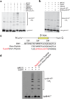
a, Fluorescence detection of cycB-NT* ubiquitination by APC/CCDH1 with UBCH10, alone or in combination with UBE2S, in the absence or presence of EMI1-SKP1 or EMI1DLZT. b, Fluorescence detection of APC-dependent UbcycB-NT* ubiquitination by UBE2S in the absence of UBCH10 and in the absence or presence of CDH1. The K0UbcycB-NT* substrate has all lysines in the Ub moiety mutated to arginines. c, Sequence alignment of the EMI1 D-box region region with Emi1 and Hsl1 D-box peptides. d, Fluorescence detection of cycB-NT* ubiquitination by APC/CCDH1 and UBCH10 in the absence or presence of the indicated D-box peptides at 100 µM.
Figure 4
Figure 4. EMI1DLZT elements synergize to mediate optimal inhibition
a, Localization of EMI1 linker and C terminus. A WD40 propeller was inserted into the Linker and at the C-terminus of EMI1FDLZT-SKP1. The 3D structures calculated contained additional densities based on the calculation of difference density maps, although the extra densities do not fully occupy the expected volume for a propeller probably due to flexibile connections. Shown are the strongest peaks in the difference maps for the Linker insertion in blue, and the C-terminal insertion in purple, relative to APC/CCDH1-EMI1-SKP1. Conformational variability was distinguished from additional densities by the fact that negative and positive difference densities are located next to each other in areas with substantial conformational flexibility, while no negative difference density can be observed for the WD40 propeller insertions. b, Human APC/CCDH1-EMI1DLZT, showing density attributed to the D-box and associated CDH1 in yellow, to the Linker in green, and to the ZBR and Tail integrated with portions of APC1 and APC4 in purple. c, Sequence alignment of the EMI1 D-box and Linker region indicating mutations. d, Fluorescence detection of APC/CCDH1- and UBCH10-dependent cycB-NT* ubiquitination either in the absence or presence of 50 nM of the indicated variant of EMI1DLZT.
Figure 5
Figure 5. NMR structure of the ZBR and identification of a surface required for inhibition
a, Solution structure of the ZBR portion of EMI1ZT as cartoon with zincs as spheres on left, and as ribbon superimposed with the RNF31 (2CT7.pdb, gold) IBR on right. b, The crystal structure of the EMI1 ZBR is shown with approximate location next to the EM structure of APC/CCDH1-EMI1DLZT at the same scale. c, Alanine scan testing roles of indicated ZBR side-chains on EMI1DZLT-mediated inhibition of cycB-NT* ubiquitination by APC/CCDH1 and UBCH10. Inhibition by wild-type EMI1DLZT and EMI1NFDL-SKP1 (lacking the ZBR and Tail) are shown for comparison. (X) denotes unfolded mutants. Mutants displaying greatest decrease in inhibitory activity are marked with purple dots those with a lesser decrease in inhibitory activity are marked with gold dots. d, Comparison of the effects of the indicated surface alanine mutants and the unfolding C409A mutant on EMI1DZLT-mediated inhibition of cycB-NT* ubiquitination by APC/CCDH1 and UBCH10. e, Structure of EMI1 ZBR, showing all side-chains mutated in Ala scan in sticks, with those causing greatest decrease in inhibitory activity in purple and a lesser decrease in inhibitory activity in gold. f, Location of Ser383 (side-chain shown as green sticks with oxygen in red) cyclin-dependendent kinase (cdk)-mediated phosphorylation site on cartoon representation of EMIZT structure with the two zinc atoms shown in spheres, the sites of Ala mutations that impair EMI1DLZT-mediated inhibition colored as in panel e. Ser383 is among three cdk sites that when phosphorylated impairs EMI1 inhibition of APC/C 49.
Figure 6
Figure 6. EMI1 C-terminal tail is a specific inhibitor of APC/C- and UBE2S–dependent ubiquitin chain formation
a, Representative fluorescence scan of raw SDS-PAGE data (SEM = Standard error of the mean) and b, curve fit for inhibition of APC/C-dependent, CDH1-independent ubiquitination of UbcycB-NT* in the presence of increasing concentrations of a peptide corresponding to the EMI1 C-terminal tail (EMI1T). SEM, n=3 c, Representative fluorescence scan of raw SDS-PAGE data and d, curve fit for inhibition of APC/C-dependent, CDH1-independent ubiquitination of Ub* in the presence of increasing concentrations of a peptide corresponding to the EMI1 C-terminal tail (EMI1T). SEM, n=3 e, Fluorescence detection of cycB-NT* ubiquitination by APC/CCDH1 by UBCH10 and UBE2S, in the absence or presence of EMI1DLZT, or increasing amounts of EMI1ZT. EMI1ZT selectively inhibits formation of HMW conjugates formed when UBE2S is included in the reaction. f, Alignment of EMI1 C-terminal sequences from the indicated organisms, and corresponding region of human EMI2 and UBE2S. Yellow - EMI1T peptide sequence.
Figure 7
Figure 7. Mechanisms of APC/C inhibition
Model for EMI1 inhibition of APC/CCDH1. EMI1’s inhibitory C-terminal domain binds multiple sites for multimodal inhibition of APC/CCDH1, blocking both substrate access to the D-box coreceptors CDH1 and APC10, and preventing Ub chain elongation by UBE2S. The N-terminal domain of EMI1 is exposed, enabling regulation by binding to other partner proteins that modulate localization, EMI1 stability,, and EMI1’s ability to bind APC/C 49.

Comment in

Similar articles

Cited by

References

    1. Deshaies RJ, Joazeiro CA. RING domain E3 ubiquitin ligases. Annu Rev Biochem. 2009;78:399–434. - PubMed
    1. Barford D. Structural insights into anaphase-promoting complex function and mechanism. Philos Trans R Soc Lond B Biol Sci. 2011;366:3605–3624. - PMC - PubMed
    1. McLean JR, Chaix D, Ohi MD, Gould KL. State of the APC/C: organization, function, and structure. Crit Rev Biochem Mol Biol. 2011;46:118–136. - PMC - PubMed
    1. da Fonseca PC, Kong EH, Zhang Z, Schreiber A, Williams MA, Morris EP, Barford D. Structures of APC/C(Cdh1) with substrates identify Cdh1 and Apc10 as the D-box co-receptor. Nature. 2011;470:274–278. - PMC - PubMed
    1. Chao WC, Kulkarni K, Zhang Z, Kong EH, Barford D. Structure of the mitotic checkpoint complex. Nature. 2012;484:208–213. - PubMed

Publication types

MeSH terms