. 2013 Jul;20(7):796-803.
doi: 10.1038/nsmb.2594. Epub 2013 May 26.

Supersite of immune vulnerability on the glycosylated face of HIV-1 envelope glycoprotein gp120

Affiliations

Supersite of immune vulnerability on the glycosylated face of HIV-1 envelope glycoprotein gp120

Leopold Kong et al. Nat Struct Mol Biol. 2013 Jul.

Abstract

A substantial proportion of the broadly neutralizing antibodies (bnAbs) identified in certain HIV-infected donors recognize glycan-dependent epitopes on HIV-1 gp120. Here we elucidate how the bnAb PGT 135 binds its Asn332 glycan-dependent epitope from its 3.1-Å crystal structure with gp120, CD4 and Fab 17b. PGT 135 interacts with glycans at Asn332, Asn392 and Asn386, using long CDR loops H1 and H3 to penetrate the glycan shield and access the gp120 protein surface. EM reveals that PGT 135 can accommodate the conformational and chemical diversity of gp120 glycans by altering its angle of engagement. Combined structural studies of PGT 135, PGT 128 and 2G12 show that this Asn332-dependent antigenic region is highly accessible and much more extensive than initially appreciated, which allows for multiple binding modes and varied angles of approach; thereby it represents a supersite of vulnerability for antibody neutralization.


Figures

Figure 1
Figure 1. Crystal structure of PGT 135 in complex with HIV-1 gp120
(a) The glycan-dependent interaction between PGT 135 and gp120 is presented in ribbon representation in the context of the gp120 ternary complex that contains a 2-domain CD4 and the Fab of CD4-induced antibody 17b. An enlarged display of the PGT 135-gp120 interaction is shown to the lower right. The CDR H1 and H3 loops (blue and red respectively) penetrate deeply through the glycan canopy to contact protein surface below while the light chain contacts the glycans using CDR loops and framework regions. (b) Buried surface on gp120 protein surface and attached glycans N332, N386 and N392 by CDR loops and framework regions of PGT 135. The values are in Å2.
Figure 2
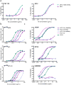
Figure 2. PGT 135 neutralization and binding
(a) Neutralization activity of PGT 135 and 2G12 were assayed against JR-FL pseudovirus in TZM-bl cells. Neutralization of WT JR-FL and of JR-FL containing V1-V2 loop mutations E168K and N188A are shown. (b) Binding of PGT 135 IgG mutants to gp120 from various strains (JRCSF, JRFL) as well as JRFLmV3 were measured by ELISA. Mutations include deletion of the CDR H1 insert “EWGDK”, alanine substitution of the CDR H2 arginine that supports the CDR H1 insert with a salt bridge, and mutation of VFMLV to AAMAA in CDR H3. Wild-type PGT 135 and 2G12 IgGs are included as controls. (c) Neutralization potency of PGT 135 mutant IgGs against JRCSF, SF162 and 92RW020. Error bars represent the standard deviation. Experiments were performed in duplicate and repeated three times.
Figure 3
Figure 3. Glycan dependency of PGT 135 binding to gp120
(a) PGT 135 interactions with gp120 glycans. The 2Fo-Fc electron density (contoured at 1σ) of N392, N332 and N386 glycans are shown along with contacting residues on PGT 135. A schematic of each glycan is shown beside the structures for the sugars that are visible in the electron density with GlcNAc as dark blue squares and mannose residues as green circles. The surface area (Å2) on each glycan moiety buried by PGT 135 is shown. (b) Interactions of PGT 135, PGT 128 and 2G12 with oligomannose glycans on a high-density microarray. The error bars represent standard deviations where n=6. (c) Glycan dependency of PGT 135 neutralization using BaL pseudoviruses produced in different cell systems. Virus produced in 293T cells contains complex, hybrid and oligomannose type glycans, whereas in 293S cells only Man5-9GlcNAc2 glycans are present. Addition of kifunensine yields only Man9GlcNAc2 glycans. NB-DNJ blocks the glycan processing at neutral glucosylated glycans. Error bars represent the standard deviation. Each experiment was performed in duplicate and repeated at least three times. (d) The effect on PGT 135 neutralization from single mutations removing glycans and His330 for several HIV-1 strains. Values are presented as fold change in IC50 of variant envelope compared to WT envelope (fold change = IC50 variant/IC50 WT). Boxes are color coded for fold change; red: >100, orange 10-100, yellow 4-9 and green <4. n.d.: not determined. a values were determined by ELISA instead of neutralization.
Figure 4
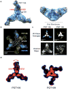
Figure 4. A gp140 trimer binds Fab PGT 135 in slightly different orientations in contrast to PGT128, which is bound in a single orientation
(a) The PGT 135-gp120 structure is shown fitted into the EM density map of PGT 135 in complex with the BG505 SOSIP.664 gp140 trimer in top and side views of the trimer. (b) 2D difference map of the two 2D class averages. The PGT128-SOSIP class average was subtracted from the PGT 135-SOSIP class average. The overlap is shown as the black gap in the trimer region. (c) Top view of the 2D class averages of the BG505 SOSIP.664 gp140 trimer in complex with PGT 135 (left) and PGT128 (right). The images contributing to the class averages were aligned using a top view back projection of an undecorated gp140 SOSIP.664 trimer. On the top, the Fabs are colored cyan and yellow for PGT 135 and PGT128, respectively. Below are variance maps of the trimer in complex with PGT 135 (left) and PGT128 (right) indicating the variability in the images that went into the 2D class averages. Brighter pixel values correspond to a greater degree of variability in that region. (d) An overlay of the 2D class average (blue outline) and the first eigenvector (orange) of the 2D PCA analysis for PGT 135 and PGT128.
Figure 5
Figure 5. Supersite of vulnerability centered on the N332 glycan
(a) Binding to the N332 glycan on gp120 for PGT 135, 2G12 and PGT 128 is shown with their respective structures superimposed on glycan N332. (b) The epitopes of PGT 135, PGT 128 and 2G12 are shown in topology representations in which cylinders are α-helices, arrows are β-strands and lines are loops. N-linked glycan sites are circled. The structures of 2G12 (PDBID: 1OP5) and PGT 128 (PDBID: 3TYG) were obtained from the PDB.
Figure 6
Figure 6. Conserved conformation of N332 glycan
(a) The N-linked glycan at N332 of gp120 has a conserved conformation in crystal structures with PGT 135 (blue) and PGT 128 (light brown). The structures are superposed on the gp120 protein. (b) Definition of A and B faces of a glycan residue. The B face (labeled in red), on which the carbons are arranged counter-clockwise around the ring, is generally more apolar than the A face (labeled in cyan), on which the carbons are arranged clockwise around the ring. (c,d) The side from which each glycan moiety interacts with PGT 135 (c) or PGT 128 (d) is defined by an arrow pointing towards the interaction (red arrow indicates B face, cyan arrow indicates A face and grey arrow indicates the side of a glycan). Each sugar primarily uses its A face, B face, or the side in the interaction with the different antibodies; their buried surface areas are estimated based on the burial on each glycan residue. The structure of PGT 128 (PDBID: 3TYG) is available from the PDB.

Comment in

References

    1. Weiss RA, et al. Neutralization of human T-lymphotropic virus type III by sera of AIDS and AIDS-risk patients. Nature. 1985;316:69–72. - PubMed
    1. Wei X, et al. Antibody neutralization and escape by HIV-1. Nature. 2003;422:307–312. - PubMed
    1. Zhang M, et al. Tracking global patterns of N-linked glycosylation site variation in highly variable viral glycoproteins: HIV, SIV, and HCV envelopes and influenza hemagglutinin. Glycobiology. 2004;14:1229–1246. - PubMed
    1. Richman DD, Wrin T, Little SJ, Petropoulos CJ. Rapid evolution of the neutralizing antibody response to HIV type 1 infection. Proc Natl Acad Sci U S A. 2003;100:4144–4149. - PMC - PubMed
    1. Scheid JF, et al. Sequence and structural convergence of broad and potent HIV antibodies that mimic CD4 binding. Science. 2011;333:1633–1637. - PMC - PubMed

Publication types

MeSH terms