. 2014 Apr 24;33(17):2264-72.
doi: 10.1038/onc.2013.157. Epub 2013 May 27.

Matrix metalloprotease 1a deficiency suppresses tumor growth and angiogenesis

Affiliations

Matrix metalloprotease 1a deficiency suppresses tumor growth and angiogenesis

C J Foley et al. Oncogene. .

Abstract

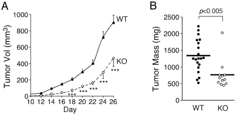
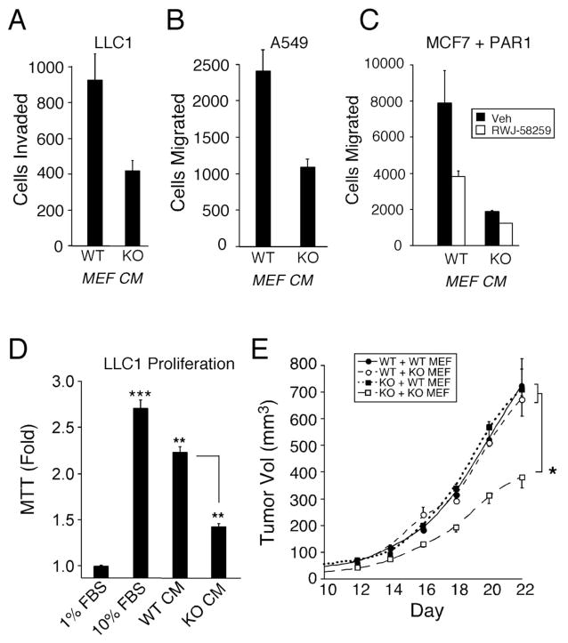
Matrix metalloprotease-1 (MMP1) is an important mediator of tumorigenesis, inflammation and tissue remodeling through its ability to degrade critical matrix components. Recent studies indicate that stromal-derived MMP1 may exert direct oncogenic activity by signaling through protease-activated receptor-1 (PAR1) in carcinoma cells; however, this has not been established in vivo. We generated an Mmp1a knockout mouse to ascertain whether stromal-derived Mmp1a affects tumor growth. Mmp1a-deficient mice are grossly normal and born in Mendelian ratios; however, deficiency of Mmp1a results in significantly decreased growth and angiogenesis of lung tumors. Coimplantation of lung cancer cells with wild-type Mmp1a(+/+) fibroblasts completely restored tumor growth in Mmp1a-deficient animals, highlighting the critical role of stromal-derived Mmp1a. Silencing of PAR1 expression in the lung carcinoma cells phenocopied stromal Mmp1a-deficiency, thus validating tumor-derived PAR1 as an Mmp1a target. Mmp1a secretion is controlled by the ability of its prodomain to facilitate autocleavage, whereas human MMP1 is efficiently secreted because of stable pro- and catalytic domain interactions. Taken together, these data demonstrate that stromal Mmp1a drives in vivo tumorigenesis and provide proof of concept that targeting the MMP1-PAR1 axis may afford effective treatments of lung cancer.


Conflict of interest statement

Conflict of Interest

The authors declare no conflict of interest.

Figures

Figure 1
Figure 1. Stromal Mmp1a-deficiency attenuates growth of lung tumors
a. Growth of Lewis lung carcinoma (LLC1) cells (2 × 10⁵) implanted subcutaneously into the abdominal fat pad of Mmp1a+/+ (n=20) or Mmp1a−/− (n=12) C57BL/6 female mice. b. Excised tumor mass at the experiment endpoint, day 26. ***p<0.001 by heteroscedastic T-Test at each time point in both genetic backgrounds.
Figure 2
Figure 2. Tumor angiogenesis is suppressed in Mmp1a-deficient animals
a. Von Willebrand Factor (vWF) immunohistochemistry on LLC1 subcutaneous tumors from wild type or Mmp1a−/− mice (40X magnification). b. Number of vWF-positive blood vessels as determined by the sum of 50 fields (40X) per tumor, n=10 per cohort. c–e. Tube formation of primary human endothelial cells (HUVECs) following 6 h stimulation with media isolated from the embryonic fibroblasts (MEFs) of wild-type or Mmp1a-deficient mice. 4X phase contrast micrographs of representative (c) cultures with corresponding quantification of (d) tubal length and (e) branch point complexity (arbitrary units). All P values were determined by heteroscedastic T-Test.
Figure 3
Figure 3. Stromal Mmp1a promotes proliferation, migration, and tumorigenesis of lung cancer
a. LLC1 chemoinvasion through type I collagen towards Mmp1a+/+ (WT) or Mmp1a−/− (KO) MEF conditioned media (CM). b. Migration of the human lung cancer cell line A549 toward WT or KO MEF conditioned media. c. Migration of MCF7 breast cancer cells ectopically expressing PAR1 towards MEF conditioned media in the absence (black) or presence (white) of the small molecule PAR1 antagonist, RWJ-58259 (3 μM). d. 96 h MTT proliferation of LLC1 cells in response to 10% FBS or MEF conditioned media. e. Tumor growth in Mmp1a+/+ (WT) or Mmp1a−/− (KO) mice co-implanted with 2 × 10⁵ LLC1 and 1 × 10⁵ Mmp1a+/+ (WT MEF) or Mmp1a−/− (KO MEF) fibroblasts (n=12–16 per cohort). * p<0.05, **p<0.005, ***p<0.001 by heteroscedastic T-Test or ANOVA followed by T-test at each time point.
Figure 4
Figure 4. Stromal Mmp1a promotes the growth of lung cancer through PAR1
a. Tumor growth following subcutaneous implantation of 200,000 shLuc control (n=10–20) versus shPAR1 (n=10) transduced LLC1 cells in Mmp1a+/+ (WT) or Mmp1a−/− (KO) mice. b. Mass of excised LLC1 tumors at the day 26 endpoint. # p=0.06, *p<0.05 by one-way ANOVA followed by T-test at each time point.
Figure 5
Figure 5. Mmp1a and MMP1 prodomain-catalytic domain interactions regulate secretion and autocatalysis
a. Structure of Mmp1a as predicted by homology modeling with human proMMP1 showing the prodomain (yellow), catalytic domain (blue), linker (green), and hemopexin domain (orange). Black arrow-zymogen activating cleavage site; arrow head-linker cleavage site resulting in loss of hemopexin domain. b. Docking of the Mmp1a prodomain (yellow) onto the catalytic domain/active site region (blue) of Mmp1a. Residues A58 and L67 in helix 2 (H2) are highlighted in red while the corresponding human residues V61 and F70, respectively are depicted in green. c. Alignment of human MMP1, Mmp1a, and Mmp1b depicting the structural motifs within the prodomain; H=helix, L=linker. Point mutations are highlighted in black. d. Secretion of Mmp1a and MMP1 prodomain mutants into the media (40 μL) of transfected HEK293T cells as determined by anti-Myc Western blot. e. MMP expression levels in cell lysates (40 μg) of transfected HEK293T cells, showing proMMP (56 kDa), active MMP (48 kDa), and hemopexin degradation product (26 kDa).
Figure 6
Figure 6. Effect of Mmp1a and MMP1 prodomains on expression of the angiogenesis factor, Cyr61
Induction of CYR61 mRNA in mouse epithelial cells (C57MG) following 24 h treatment with MMP-transfected Cos7 CM as described in the Methods. Data represent means ± SE of triplicate experiments.

References

    1. Boström P, Söderström M, Vahlberg T, Söderström K-O, Roberts PJ, Carpén O, et al. MMP-1 expression has an independent prognostic value in breast cancer. BMC Cancer. 2011;11:348. - PMC - PubMed
    1. Smith V, Wirth GJ, Fiebig HH, Burger AM. Tissue microarrays of human tumor xenografts: characterization of proteins involved in migration and angiogenesis for applications in the development of targeted anticancer agents. Cancer Genomics Proteomics. 2008;5:263–273. - PMC - PubMed
    1. Kanamori Y, Matsushima M, Minaguchi T, Kobayashi K, Sagae S, Kudo R, et al. Correlation between expression of the matrix metalloproteinase-1 gene in ovarian cancers and an insertion/deletion polymorphism in its promoter region. Cancer Research. 1999;59:4225–4227. - PubMed
    1. Nikkola J, Vihinen P, Vlaykova T, Hahka-Kemppinen M, Kähäri V-M, Pyrhönen S. High expression levels of collagenase-1 and stromelysin-1 correlate with shorter disease-free survival in human metastatic melanoma. Int J Cancer. 2002;97:432–438. - PubMed
    1. Murray GI, Duncan ME, O’Neil P, Melvin WT, Fothergill JE. Matrix metalloproteinase-1 is associated with poor prognosis in colorectal cancer. Nat Med. 1996;2:461–462. - PubMed

Publication types

MeSH terms