. 2013 Jun 20;498(7454):318-24.
doi: 10.1038/nature12296. Epub 2013 May 24.

The linear ubiquitin-specific deubiquitinase gumby regulates angiogenesis

Affiliations

The linear ubiquitin-specific deubiquitinase gumby regulates angiogenesis

Elena Rivkin et al. Nature. .

Abstract

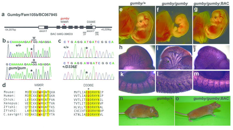
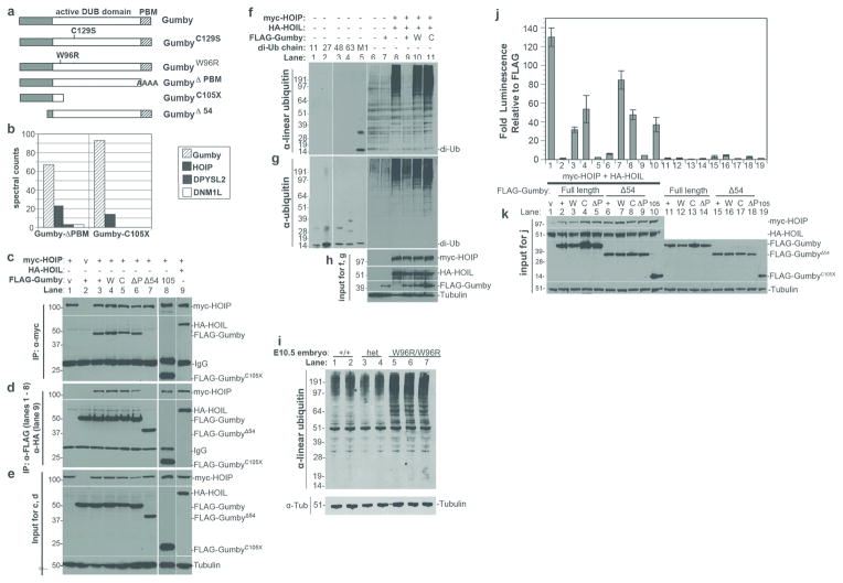
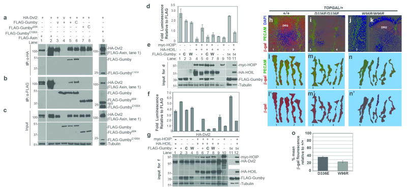
A complex interaction of signalling events, including the Wnt pathway, regulates sprouting of blood vessels from pre-existing vasculature during angiogenesis. Here we show that two distinct mutations in the (uro)chordate-specific gumby (also called Fam105b) gene cause an embryonic angiogenic phenotype in gumby mice. Gumby interacts with disheveled 2 (DVL2), is expressed in canonical Wnt-responsive endothelial cells and encodes an ovarian tumour domain class of deubiquitinase that specifically cleaves linear ubiquitin linkages. A crystal structure of gumby in complex with linear diubiquitin reveals how the identified mutations adversely affect substrate binding and catalytic function in line with the severity of their angiogenic phenotypes. Gumby interacts with HOIP (also called RNF31), a key component of the linear ubiquitin assembly complex, and decreases linear ubiquitination and activation of NF-κB-dependent transcription. This work provides support for the biological importance of linear (de)ubiquitination in angiogenesis, craniofacial and neural development and in modulating Wnt signalling.


Conflict of interest statement

The authors declare no competing financial interests.

Figures

Figure 1
Figure 1. Identification of the gumby (GumW96R) causative mutation and the new GumD336E allele
a, Schematic diagram of the Gumby/Fam105b/BC067945 gene and BAC bMQ-396D3. Primers 7F and 7R used to identify mice carrying GumD336E flank exon 7. Sequencing traces from b, +/+ and gumby/gumby (gum/gum) and c, +/+ and GumD336E/+ (+/D336E) mice. d, Amino acid sequence alignment spanning GumW96R and GumD336E mutations. Trp96 and Asp336 are shown in red, marked with asterisks. Yellow highlights interspecies amino acid sequence identity. (e–o) BAC rescue of lethality and vascular abnormalities of gumby/gumby mice. Morphological appearance of e, gumby/+, f, gumby/gumby, and g, gumby/gumby embryos carrying the BAC transgene (gumby/gumby;BAC) at E14.0. h–m, PECAM-1 immunohistochemistry of h, k, gumby/+, i, l, gumby/gumby and j, m, gumby/gumby;BAC embryos at E10.5. h–j, cranial vasculature and k–m, trunk vasculature shown at the forelimb bud level. Arrow indicates dilated cranial vessels; arrowheads highlight stunted branches in the trunk. n, adult gumby/+ and o, gumby/gumby;BAC littermates.
Figure 2
Figure 2. Analyses of vascular phenotypes and Gumby expression
a–f, Whole mount anti-PECAM-1 immunofluorescence showing cranial vasculature of E10.5 a, a′, b, +/+, c, c′, d, GumW96R/GumW96R (W96R) and e, e′, f, GumD336E/GumD336E (D336E) embryos. a, c, e, Composite whole head images. a′, c′, e′, Secondary and tertiary vessels are marked in purple and green, respectively. b, d, f, Analyses of cranial vessel dilation of intercranial artery (ICA). White bars indicate the areas of the ICA (h) and first branch (i) measured. g, Quantification of secondary and tertiary vessels branching off the primary ICA per side of head. h, Quantification of h, ICA and i, first ICA branch diameters. * indicates statistical significance relative to +/+ embryos (n = 3 or 6, p < 0.05, Student’s t-test). j–l, RNA in situ hybridization of E10.5 +/+ embryos detects Gumby RNA in the vasculature. Magnified views from k, head and l, trunk. An intersomitic vessel (ISV) is marked with an arrow. m–p, Immunofluorescence with anti-PECAM-1 (green) and anti-Gumby (red) antibodies detects Gumby in endothelial cells in cross-sections of the trunk in +/+ E11.5 embryos. n High magnification of boxed area in m. Gumby protein is enriched near presumptive tips of vessels (arrows) and vascular buds (arrowheads).
Figure 3
Figure 3. Structural and biochemical analysis of Gumby
a, Ribbons view of apo-Gumby (green) superimposed on OTUB1 (PDB 4DDG) (grey). Inset shows catalytic triad. b, Cleavage specificity towards di-ubiquitin chains c, Ribbons view of the Gumby-linear di-ubiquitin complex. d, Peel-away surface views of linear di-ubiquitin (top) bound to Gumby (bottom). Domains colored as in c, with residues involved in inter-molecular contacts colored according to the domains contacted. Catalytic triad and mutant positions labeled green and red respectively. e, Active site comparison of Gumby-di-ubiquitin and Gumby-mono ubiquitin complexes. A productive orientation of His339 is observed only in the Gumby-di-ubiquitin complex.
Figure 4
Figure 4. Gumby interacts with HOIP/RNF31 and counteracts LUBAC activity
a, FLAG-tagged Gumby constructs used. b, Representative mass spectromety results for FLAG-tagged GumbyΔPBM and GumbyC105X immunoprecipitations. HOIP was identified as the most significant interacting protein in HEK293 cells expressing FLAG-tagged GumbyΔPBM or GumbyC105X. Significance threshold was set at a protein probability of greater than 90% (determined using SAINT analysis), less than 10% project frequency and a minimum of 3 peptides. c–e, Immunoprecipitation coupled to immunoblotting using FLAG-Gumby expression constructs, HA-HOIL and myc-HOIP determined that HOIP interacts with wild type Gumby (+), GumbyC129S (C), GumbyΔPBM (P) and GumbyC105X (105) proteins, but not GumbyΔ54 (54). c, Immunoblot of immunoprecipitation using anti-myc antibody for myc-HOIP. d, Immunoblot of immunoprecipitation using anti-FLAG antibody for FLAG-tagged Gumby constructs. e, inputs for immunoprecipitation experiments. f–h, Immunoblot using anti-linear ubiquitin antibody detected decreased levels of linear ubiquitinated proteins in HEK293T cells, when Gumby, but not GumbyC129S or GumbyW96R, was co-expressed with HOIL and HOIP. The anti-linear ubiquitin antibody recognizes linear diubiquitin (M1), but not control diubiquitin chains K11 (11), K48 (48), K63 (63). g, Equivalent levels of ubiquitination were detected in HOIL-HOIP co-expressing cells by anti-pan-ubiquitin antibody. h, Inputs for f and g. i, Immunoblot with anti-linear ubiquitin antibody detects increased levels of linear ubiquitinated protein in individual E10.5 GumW96R/GumW96R embryos (W96R/W96R) relative to +/+ and /+ (het) littermates. j, Catalytically active Gumby counteracts HOIP-HOIL dependent stimulation of luciferase expression from an NF-κB reporter in HEK293T cells. Wildtype, GumbyΔ54, GumbyΔPBM or GumbyΔ54ΔPBM fully suppress this stimulation. Data are presented as means +/− s.e.m. (n=3; P<0.05, Student’s t-test). k, Immunoblot of inputs for luciferase assays.
Figure 5
Figure 5. Gumby interacts with Dvl2 and can modulate Wnt signaling
a, Immunoprecipitation (IP) with anti-HA antibody of HA-tagged Dvl2 (HA-Dvl2) recovered FLAG-tagged Gumby, GumbyC129S (C), GumbyC105X (105) and FLAG-Axin (positive control), but not GumbyΔ54 (54). FLAG-vector is designated as “v”. b, In immunoprecipitation with anti-FLAG antibody, all FLAG-Gumby constructs except for FLAG-GumbyΔ54 recovered HA-Dvl2. c, Input amounts and tubulin levels. d, f, In assays for Wnt3a activation of luciferase expression from the TOPFLASH reporter performed in d,e, the absence or f, g, presence of HA-Dvl2, FLAG-Gumby enhanced TOPFLASH expression more than FLAG-GumbyW96R (W) and FLAG-GumbyC129S. HA-HOIL and myc-HOIP co-expression inhibited TOPFLASH. Gumby, but not GumbyC129S or GumbyW96R, reversed HOIL-HOIP dependent inhibition. e, g, Immunoblots show inputs for luciferase assays in d and f, respectively. Data are presented as means +/− s.e.m. n=3 or more. (P<0.05, Student’s t-test). h–o Analysis of β-galactosidase (β-gal) expression in intersomitic vessels (ISVs) of E10.5 embryos. h, Co-immunofluorescence with anti-β-gal (red) and anti-PECAM-1 (green) antibodies detects high βgal within ISVs and dorsal root ganglia (DRG) of +/+;TOPGAL/+ embryos. In i, GumD336E/GumD336E;TOPGAL/+ and j,k, GumW96R/GumW96R;TOPGAL+ embryos, β-gal signal is lower in ISVs and in DRG. k–m′, representative traces of ISVs scored for β-gal fluorescence intensity are shown for l,l′, +/+;TOPGAL/+, m,m′, GumD336E/GumD336E;TOPGAL/+ and n,n′, GumW96R/GumW96R;TOPGAL+ embryos. Numbers in k–m′ mark specific ISVs traced in h, i and j. n, Graph of percentage mean fluorescence in ISVs of GumD336E/GumD336E;TOPGAL/+ (D336E) and GumW96R/GumW96R;TOPGAL+ (W96R) embryos. (36.6 +/− 1.8% and 24.0 +/− 2.8%, respectively). Data are presented as means +/− s.e.m. Effect of Gumby genotype on anti-β-gal mean fluorescence intensity was highly significant (Kruskal-Wallis test (k=3); H=43.69; p<0.0001).

Comment in

References

    1. Risau W. Mechanisms of angiogenesis. Nature. 1997;386(6626):671–674. - PubMed
    1. Logan CY, Nusse R. The Wnt signaling pathway in development and disease. Annu Rev Cell Dev Biol. 2004;20:781–810. - PubMed
    1. Liebner S, Plate KH. Differentiation of the brain vasculature: the answer came blowing by the Wnt. Journal of angiogenesis research. 2010;2:1. - PMC - PubMed
    1. Zerlin M, Julius MA, Kitajewski J. Wnt/Frizzled signaling in angiogenesis. Angiogenesis. 2008;11(1):63–69. - PubMed
    1. Daneman R, et al. Wnt/beta-catenin signaling is required for CNS, but not non-CNS, angiogenesis. Proc Natl Acad Sci U S A. 2009;106(2):641–646. - PMC - PubMed

Publication types

MeSH terms