. 2013 Sep;11(9):1061-1071.
doi: 10.1158/1541-7786.MCR-13-0111. Epub 2013 May 24.

A novel germline mutation in BAP1 predisposes to familial clear-cell renal cell carcinoma

Affiliations

A novel germline mutation in BAP1 predisposes to familial clear-cell renal cell carcinoma

Megan N Farley et al. Mol Cancer Res. 2013 Sep.

Abstract

Renal cell carcinoma (RCC) clusters in some families. Familial RCC arises from mutations in several genes, including the von Hippel-Lindau (VHL) tumor suppressor, which is also mutated in sporadic RCC. However, a significant percentage of familial RCC remains unexplained. Recently, we discovered that the BRCA1-associated protein-1 (BAP1) gene is mutated in sporadic RCC. The BAP1 gene encodes a nuclear deubiquitinase and appears to be a classic two-hit tumor suppressor gene. Somatic BAP1 mutations are associated with high-grade, clear-cell RCC (ccRCC) and poor patient outcomes. To determine whether BAP1 predisposes to familial RCC, the BAP1 gene was sequenced in 83 unrelated probands with unexplained familial RCC. Interestingly, a novel variant (c.41T>A; p.L14H) was uncovered that cosegregated with the RCC phenotype. The p.L14H variant targets a highly conserved residue in the catalytic domain, which is frequently targeted by missense mutations. The family with the novel BAP1 variant was characterized by early-onset ccRCC, occasionally of high Fuhrman grade, and lacked other features that typify VHL syndrome. These findings suggest that BAP1 is an early-onset familial RCC predisposing gene.

Implications: BAP1 mutations may drive tumor development in a subset of patients with inherited renal cell cancer.


Figures

Figure 1
Figure 1. Family NCI-1326: an early onset, aggressive form of bilateral, multifocal solid and cystic clear cell kidney cancer
Pedigree from familial renal cancer kindred NCI-1326. The proband IV:1 was initially diagnosed with kidney cancer at the age of 44. Individual IV:4 was diagnosed with RCC at 40 and individual IV:6 died of metastatic RCC at age 36. Individuals II:1 and III:4 died of metastatic RCC at 48 and 58 years of age, respectively. Two individuals with a history of RCC for whom samples were available (IV:1 and IV:4) had a germline BAP1 variant. Two other individuals (IV:3 and IV:5) who were screened with abdominal imaging and were found to have no evidence of RCC, were negative for the germline BAP1 gene variant. Individual IV:2 (omitted from pedigree) is the spouse of IV:1.
Figure 2
Figure 2. Bilateral, multifocal cysts and solid kidney cancer in proband IV:1 from Family NCI-1326
Axial abdominal computed tomography (CT) scans from the proband (IV:1, NCI-1326 kindred) following right radical nephrectomy showing multifocal left renal lesions, indicated by arrows in A-D. Subsequently, at the age of 46 the individual underwent the first of three left partial nephrectomies, for the surgical removal of a 3 cm ccRCC (Fuhrman grade III), a 1.5 cm ccRCC (Fuhrman grade III) and a 2 cm atypical cyst with clear cell lining.
Figure 3
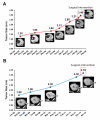
Figure 3. Rapid growth of renal tumors in proband IV:1 from Family NCI-1326
Rapid growth rate of proband IV:1 renal tumors prior to second (A), and third left partial nephrectomies (B).
Figure 4
Figure 4. Recurrent multifocal cysts and solid kidney cancer in proband IV:1 from Family NCI-1326
(A-C) Second left partial nephrectomy from proband (IV:1, NCI-1326 kindred). Axial abdominal computed tomography (CT) scan (A), and histology slides from removed lesions, a ccRCC (B) and an atypical renal cyst with clear cell lining (arrows) (C).
Figure 5
Figure 5. Cosegregation and LOH studies of BAP1 variant in Family NCI-1326
(A) Sequence chromatogram for proband IV:1 and affected individual IV:4 with the c.41T>A (p.L14H) BAP1 variant. Unaffected individuals IV:3 and IV:5 were negative for the BAP1 variant. (B) Sequence chromatograms of renal tumors from proband IV:1 displaying loss of heterozygosity (LOH) at the BAP1 locus for 2 different regions from one tumor (Tumors 1a and 1b) and mutant allele enrichment in another tumor (Tumor 3). Sequence chromatogram of individual IV:4 tumor showing LOH. Nx, nephrectomy.
Figure 6
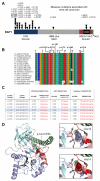
Figure 6. Analysis of the novel p.L14H BAP1 variant
(A) Schematic of BAP1 protein showing the position of the novel p.L14H missense variant (gray triangle) in comparison to known BAP1 missense mutations associated with sporadic RCC (black triangles). Data compiled from Peña-Llopis et al., Hakimi et al., COSMIC and KIRC (TCGA) (11, 21, 24). UCH, ubiquitin C-terminal hydrolase domain (blue); HBM, HCF-1-binding motif (yellow); ULD, Uch37-like domain (black); BRCA1, putative BRCA1-interacting domain (red); NLS, nuclear localization signal (green). (B) BAP1 amino acid conservation across species assessed with BioEdit’s ClustalW multiple alignment function. Protein sequences are from UniProt (Q92560, H9G0D9, F6TYN2, E2R9Z2, D3ZH56, Q99PU7, G1PS27, F6SMM8, F6RI15, Q5F3N6, Q52L14, H2UEV1, A1L2G3, Q7K5N4, Q17N72) and Ensembl (ENSPTRP00000025898) (33). (C) In silico predicted effects of the novel p.L14H missense variant and the surrounding sporadic RCC-associated missense mutations assessed with PROVEAN, SIFT and PolyPhen-2 prediction tools (19, 25, 26). (D) BAP1 structure model. Left Panel: Cartoon depiction of the BAP1 UCH domain (purple) noting p.Leu14 (red sphere) involved in organizing a flexible crossover loop and other flexible portions of the domain (salmon) that order upon ubiquitin substrate (cyan) binding. Uch37-like domain (ULD) is shown in green. Right Upper Panel: Zoom-in of wild type BAP1 leucine 14 residue with atom radii depicted in dots interacting with surrounding residues (side chains within 4 Å displayed in stick). Right Lower Panel: Zoom-in of BAP1 histidine 14 mutant with atom radii (dots) revealing clashes with surrounding residues.
Figure 7
Figure 7. Loss of BAP1 protein in renal tumor from individual IV:4
Low power view of renal tumor from individual IV:4. The tumor involves the medullary area of the kidney and shows negative staining for BAP1. Note the pelvic transitional epithelium that stains positive for BAP1 (*) as positive internal control (150X). Insert A: High power image showing the renal tumor with negative BAP1 immunohistochemical staining (200X). Insert B: High power image of BAP1 immunohistochemical staining showing pelvic transitional epithelium with positive nuclear staining as a positive internal control (250X).

References

    1. Pavlovich CP, Schmidt LS. Searching for the hereditary causes of renal-cell carcinoma. Nat Rev Cancer. 2004;4:381–93. - PubMed
    1. Linehan WM. Genetic basis of kidney cancer: role of genomics for the development of disease-based therapeutics. Genome Res. 2012;22:2089–100. - PMC - PubMed
    1. Malinoc A, Sullivan M, Wiech T, Kurt Werner S, Jilg C, Straeter J, et al. Biallelic inactivation of the SDHC gene in renal carcinoma associated with paraganglioma syndrome type 3. Endo Relat Ca. 2012;19:283–90. - PubMed
    1. Ricketts CJ, Shuch B, Vocke CD, Metwalli AR, Bratslavsky G, Middelton L, et al. Succinate dehydrogenase kidney cancer: an aggressive example of the Warburg effect in cancer. J Urol. 2012;188:2063–71. - PMC - PubMed
    1. Eletr ZM, Wilkinson KD. An emerging model for BAP1’s role in regulating cell cycle progression. Cell Biochem Biophys. 2011;60:3–11. - PMC - PubMed

Publication types

MeSH terms

Supplementary concepts