

Juvenile cellular pleomorphic adenoma
- PMID: 23709529
- PMCID: PMC3670021
- DOI: 10.1136/bcr-2012-007641
Juvenile cellular pleomorphic adenoma
Abstract
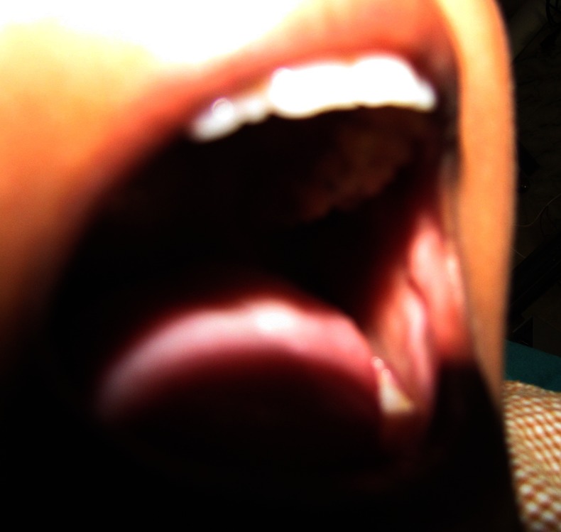
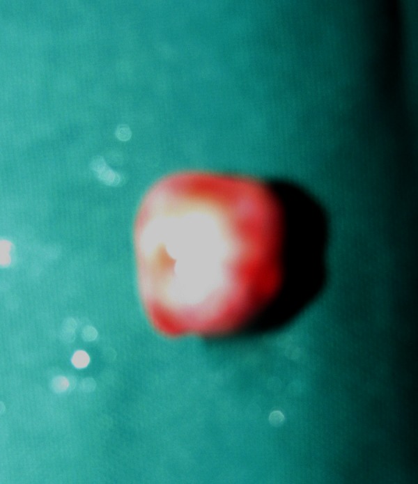
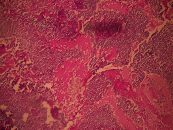
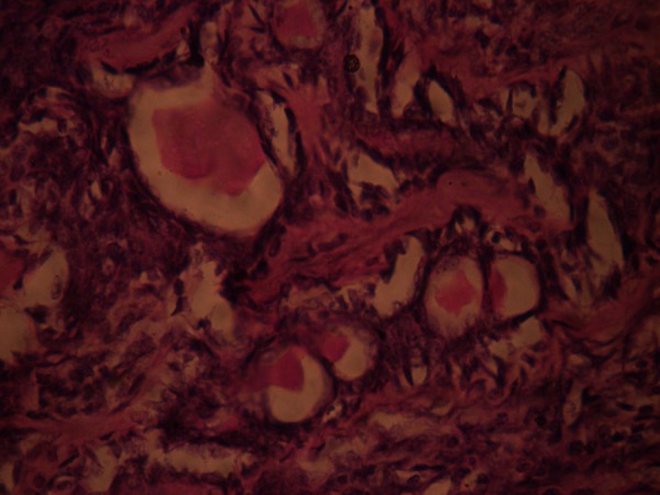
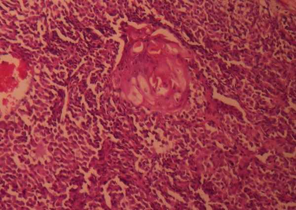
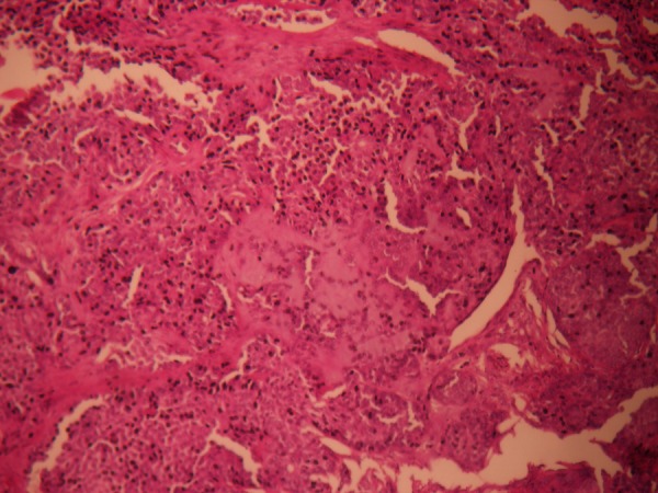
Salivary gland tumours are rare in children and when they do arise, they preferentially affect major salivary glands with sporadic incidence in minor salivary glands. The mucosa of the cheek is an uncommon site of occurrence for intraoral pleomorphic adenoma and most of these cases have been reported in adults. Histologically, it shows a highly variable morphology because of interplay between epithelial and mesenchymal (myxoid, hyaline, chondroid, osseous) elements which arise from same cell clone, which may be a myoepithelial or ductal reserve cell. Here we report a rare case of juvenile pleomorphic adenoma of the cheek in a 12-year-old girl with a predominant epithelial component histologically. Relevant studies are discussed with a focus on its cytology and cytogenetics.
Figures


References
-
- Ellis GL, Auclair PL. Atlas of tumor pathology; tumors of the salivary glands. Washington, DC: Armed Forces Institute of Pathology, 1996; third series. Fascicle 17
-
- Gnepp DR. Salivary gland (major and minor) and lacrimal gland. In: Gnepp DR. Diagnostic surgical pathology of the head and neck. 2nd edn Philadelphia: WB Saunders, 2009:434–49
-
- Lucas RB. Pathology of tumors of oral tissues. 4th edn. Edinburgh: Churchill Livingstone, 1984:298–9
-
- Krolls SO, Trodahl JN, Boyers RC. Salivary gland lesions in children. A survey of 430 cases. Cancer 1972;2013:459–69 - PubMed
Publication types
MeSH terms
LinkOut - more resources
Full Text Sources
Other Literature Sources
Medical