. 2013 May 24;2(3):e000048.
doi: 10.1161/JAHA.113.000048.

FAMP, a novel apoA-I mimetic peptide, suppresses aortic plaque formation through promotion of biological HDL function in ApoE-deficient mice

Affiliations

FAMP, a novel apoA-I mimetic peptide, suppresses aortic plaque formation through promotion of biological HDL function in ApoE-deficient mice

Yoshinari Uehara et al. J Am Heart Assoc. .

Abstract

Background: Apolipoprotein (apo) A-I is a major high-density lipoprotein (HDL) protein that causes cholesterol efflux from peripheral cells through the ATP-binding cassette transporter A1 (ABCA1), thus generating HDL and reversing the macrophage foam cell phenotype. Pre-β1 HDL is the smallest subfraction of HDL, which is believed to represent newly formed HDL, and it is the most active acceptor of free cholesterol. Furthermore it has a possible protective function against cardiovascular disease (CVD). We developed a novel apoA-I mimetic peptide without phospholipids (Fukuoka University ApoA-I Mimetic Peptide, FAMP).

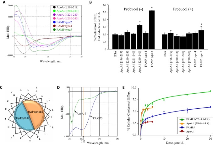
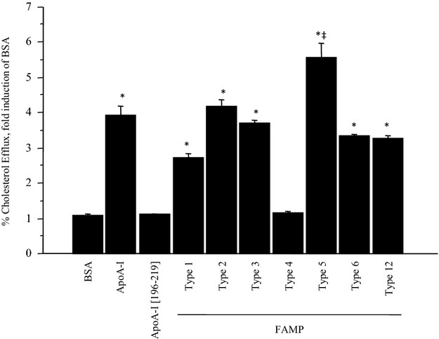
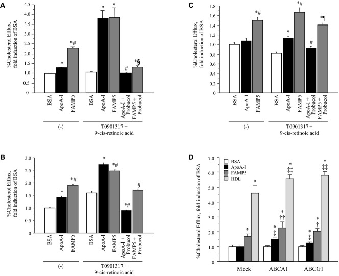
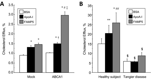
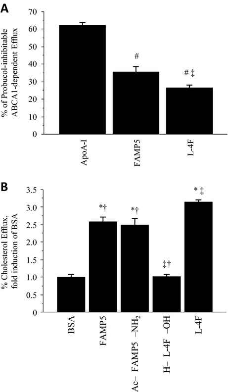
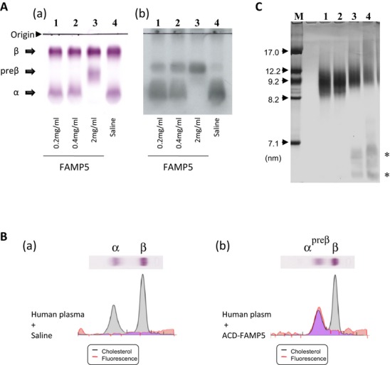
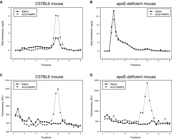
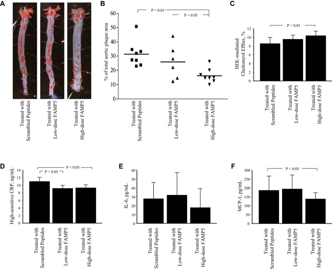
Methods and results: FAMP type 5 (FAMP5) had a high capacity for cholesterol efflux from A172 cells and mouse and human macrophages in vitro, and the efflux was mainly dependent on ABCA1 transporter. Incubation of FAMP5 with human HDL or whole plasma generated small HDL particles, and charged apoA-I-rich particles migrated as pre-β HDL on agarose gel electrophoresis. Sixteen weeks of treatment with FAMP5 significantly suppressed aortic plaque formation (scrambled FAMP, 31.3 ± 8.9% versus high-dose FAMP5, 16.2 ± 5.0%; P<0.01) and plasma C-reactive protein and monocyte chemoattractant protein-1 in apoE-deficient mice fed a high-fat diet. In addition, it significantly enhanced HDL-mediated cholesterol efflux capacity from the mice.

Conclusions: A newly developed apoA-I mimetic peptide, FAMP, has an antiatherosclerotic effect through the enhancement of the biological function of HDL. FAMP may have significant atheroprotective potential and prove to be a new therapeutic tool for CVD.

Keywords: ATP‐binding cassette transporters; HDL particle size; apolipoproteins; peptides; pre‐β HDL.


Figures

Figure 1.
Figure 1.
Characteristics of human apoA‐I fragments and apoA‐I mimetic peptides. A, Circular dichroism (CD) spectra of human apoA‐I fragments and FAMP5 in water. The apoA‐I peptide fragment consisting of amino acids 210 to 240 was the only fragment in the region of apoA‐I that formed an α‐helical conformation. FAMP2 and FAMP5, but not FAMP4, formed strong α‐helical conformations. B, Effects of various human apoA‐I fragments and FAMPs on cellular cholesterol efflux in A172 human cells. Cellular cholesterol efflux mediated by apoA‐I fragments and FAMP4 and FAMP5 were measured in human A172 cells with or without probucol (10 μmol/L) in the presence of T0901317 (5 μmol/L) and 9‐cis‐retinoic acid (9cisRA; 5 μmol/L). The experiments were performed for 4 hours in the presence or absence of 20 μg/mL apoA‐I fragments, or FAMPs (n=3 to 5 for each group). C, Helix wheel representation of FAMP5. D, CD spectra of human apoA‐I and FAMP5 in buffer with 3 mol/L guanidine hydrochloride, 10 mmol/L Tris, and 5 mmol/L dithiothreitol (DTT). E, Dose‐dependent increases on a molar basis in human apoA‐I‐ and FAMP5‐mediated cellular cholesterol efflux for 4 hours are shown in the presence or absence of 5 μmol/L of T0901317 or 9‐cis‐retinoic acid in A172 cells (n=4 each). Values are mean±SD. *P<0.01 vs BSA group. FAMP indicates Fukuoka University apoA‐I mimetic peptide; apoA‐I, apolipoprotein A‐I; BSA, bovine serum albumin.
Figure 2.
Figure 2.
Effects of candidate apoA‐I mimetic peptides on cholesterol efflux in A172 cells. Cellular cholesterol efflux mediated by novel apoA‐I mimetic peptides (FAMPs) were measured in A172 cells. The cells were treated with T0901317 (5 μmol/L) and 9‐cis‐retinoic acid (5 μmol/L) in the presence or absence of 20 μg/mL human apoA‐I, apoA‐I fragments, or FAMPs for 4 hours (n=4 to 5 for each group). Values are mean±SD. *P<0.01 vs BSA; ‡P<0.01 vs apoA‐I. FAMP indicates Fukuoka University apoA‐I mimetic peptide; apoA‐I, apolipoprotein A‐I; BSA, bovine serum albumin.
Figure 3.
Figure 3.
Effects of FAMP5 on cellular cholesterol efflux. ApoA‐I‐ and FAMP5‐mediated cholesterol effluxes were measured in A172 cells (A), mouse peritoneal macrophages (B), and human monocyte‐derived macrophages (C) in the presence or absence of T0901317 (5 μmol/L), 9‐cis‐retinoic acid (5 μmol/L), and probucol (10 μmol/L). All experiments were performed for 4 hours in the presence or absence of 20 μg/mL human apoA‐I or FAMP5 (n=4 to 5 for each group). D, COS‐7 cells were transiently transfected with the empty vector (mock) or with human ABCA1 and ABCG1 cDNA, and cholesterol efflux was measured after incubation with 20 μg/mL apoA‐I, FAMP5, or HDL. All experiments were performed for 4 hours (n=6 to 7 for each group). Values are mean±SD. *P<0.01 vs BSA; #P<0.01 vs apoA‐I; §P<0.01 vs FAMP5; ¶P<0.05 vs FAMP5; †P<0.05 vs FAMP5 in mock; ††P<0.01 vs FAMP5 in mock; ‡P<0.01 vs apoA‐I in mock; ‡‡P<0.01 vs HDL in mock. FAMP indicates Fukuoka University apoA‐I mimetic peptide; apoA‐I, apolipoprotein A‐I; BSA, bovine serum albumin; ABCA1, ATP‐binding cassette transporter A1; ABCG1, ATP‐binding cassette transporter G1; HDL, high‐density lipoprotein.
Figure 4.
Figure 4.
Effects of FAMP5 on cellular cholesterol efflux in ABCA1‐overexpressing and ‐deficient cells. A, Cultivated CHO‐ldlA7 cells were transfected with the empty vector (mock) or human ABCA1 cDNA, and cholesterol efflux was measured (n=6 for each group). All experiments were performed for 4 hours in the presence or absence of 20 μg/mL human apoA‐I or FAMP5. B, Cholesterol efflux was measured in monocyte‐derived macrophages from a healthy subject and a Tangier disease patient. All experiments were performed for 4 hours in the presence or absence of 20 μg/mL human apoA‐I or FAMP5 (n=4 for each group). Values are mean±SD. *P<0.01 vs BSA; **P<0.05 vs BSA; #P<0.01 vs apoA‐I; ##P<0.05 vs apoA‐I; †P<0.05 vs mock; ‡P<0.01 vs mock; §P<0.01 vs healthy subject. FAMP indicates Fukuoka University apoA‐I mimetic peptide; ABCA1, ATP‐binding cassette transporter A1; CHO, Chinese hamster ovary; apoA‐I, apolipoprotein A‐I; BSA, bovine serum albumin.
Figure 5.
Figure 5.
Effects of FAMP5 and other apoA‐I mimetic peptides on cellular cholesterol efflux in A172 human cells. A, Graph represents percent of probucol (10 μmol/L)–inhibitable ABCA1‐dependent cholesterol efflux mediated by apoA‐I, FAMP5, or L‐4F measured in A172 cells. B, Effects of H‐FAMP5‐OH (FAMP5), Ac‐FAMP5‐NH2, Ac‐L‐4F‐NH2 (L‐4F), and H‐L‐4F‐OH on cellular cholesterol efflux in A172 cells. The experiments were performed for 4 hours in the presence or absence of 20 μg/mL of peptides (n=5 for each group). Values are mean±SD. *P<0.01 vs BSA; #P<0.01 vs apoA‐I; ‡P<0.01 vs FAMP5; †P<0.01 vs L‐4F. FAMP indicates Fukuoka University apoA‐I mimetic peptide; ABCA1, ATP‐binding cassette transporter A1; apoA‐I, apolipoprotein A‐I; BSA, bovine serum albumin.
Figure 6.
Figure 6.
Blood clearance and influence of hemolysis on FAMP5. A, Pharmacokinetics of FAMP5 in C57BL6 and apoE‐deficient mice. Both C57BL6 (n=3) and apoE‐deficient (n=3) mice were intravenously administered 10 mg/kg of fluorescence‐labeled ACD‐FAMP5. Fifteen minutes after ACD‐FAMP5 injection, plasma fluorescence signals peaked and then became attenuated. B, Effect of FAMP5 on red cell hemolysis. Various concentrations of FAMP5 were incubated with red blood cells for 10 minutes at 37°C. Hemolysis was measured as hemoglobin content at an optical density at 540 nm of the supernatant. Hemolysis was expressed as a percentage of the Triton X‐100 lysis. Values are mean±SD. FAMP indicates Fukuoka University apoA‐I mimetic peptide; apoE, apolipoprotein E; ACD, acridone.
Figure 7.
Figure 7.
Effects of FAMP on pre‐β HDL formation in vitro. A, FAMP5 (0.2 to 2.0 mg/mL) was incubated with human whole plasma at 37°C for 60 minutes, and the plasma lipoprotein profiles were analyzed by agarose gel electrophoresis (a) and Western blotting with anti‐apoA‐I (b). FAMP5 promoted the migration of apoA‐I to the pre‐β HDL position in a dose‐dependent manner. B, Saline (a) or fluorescence‐labeled ACD‐FAMP5 2.0 mg/mL (b) was incubated with human plasma at 37°C for 60 minutes. A fluorescence signal on the agarose gel under UV light was detected in samples incubated with ACD‐FAMP5. C, FAMP5 was incubated with human HDL (2.22 mg/mL) at 37°C for 60 minutes and subjected to 5% to 20% gradient polyacrylamide gel electrophoresis. After electrophoresis, the gels were stained for proteins with CBB. Incubation with FAMP5 generated small HDL particles (*), such as pre‐β1 HDL‐like particles. Lane 1, saline; lane 2, 50 μg/mL FAMP5; lane 3, 250 μg/mL FAMP5; lane 4, 500 μg/mL FAMP5. FAMP indicates Fukuoka University apoA‐I mimetic peptide; apoA‐I, apolipoprotein A‐I; ACD, acridone; HDL, high‐density lipoprotein; CBB, Coomassie Brilliant Blue.
Figure 8.
Figure 8.
FPLC analyses of lipoprotein profiles in C57BL6 and apoE−/− mice. Mice were intraperitoneally injected with saline (closed circles) or ACD‐FAMP5 (50 mg/kg body weight), and plasma samples were collected after 24 hours. Equal volumes of plasma samples were pooled from C57BL6 (A, C) and ApoE−/− (B, D) mice, and the fractionated lipoproteins were analyzed for cholesterol (A, B) and fluorescent signals (C, D). Fractions 3 to 7 contain VLDL, fractions 8 to 16 contain LDL, and fractions 17 to 24 contain HDL. FPLC indicates fast protein liquid chromatography; FAMP, Fukuoka University apoA‐I mimetic peptide; apoE, apolipoprotein E; ACD, acridone; VLDL, very‐low‐density lipoprotein; LDL, low‐density lipoprotein; HDL, high‐density lipoprotein; RFU, relative fluorescent units.
Figure 9.
Figure 9.
Suppressive effect of FAMP on atherosclerotic lesion formation in apoE−/− mice fed a high‐fat diet. ApoE−/− mice fed a high‐fat diet (0.5% cholesterol and 10% fat) were intraperitoneally treated 3 times per week with scrambled FAMP (n=8), low‐dose FAMP5 (10 mg/kg body weight; n=7), or high‐dose FAMP5 (50 mg/kg body weight; n=8). After 16 weeks, whole aortas were collected and stained with Oil Red O (A). The extent of atherosclerosis is expressed as the percent of the lesion area extending from the ascending aorta to the abdominal bifurcation (B). The biological functioning of HDL was measured as ex vivo cholesterol efflux from RAW264 mouse macrophages mediated with HDL collected from FAMP5‐treated apoE−/− mice (C). Plasma high‐sensitive C‐reactive protein (CRP) level (D), plasma interleukin 6 (IL‐6) level (E), and plasma monocyte chemoattractant protein‐1 (MCP‐1) level (F) were measured in apoE−/− mice. Values are mean±SD. FAMP, Fukuoka University apoA‐I mimetic peptide; apoE, apolipoprotein E; HDL, high‐density lipoprotein.

Similar articles

Cited by

References

    1. von Eckardstein A, Nofer JR, Assmann G. High density lipoproteins and arteriosclerosis. Role of cholesterol efflux and reverse cholesterol transport. Arterioscler Thromb Vasc Biol. 2001; 21:13-27 - PubMed
    1. Yancey PG, Bortnick AE, Kellner‐Weibel G, de la Llera‐Moya M, Phillips MC, Rothblat GH. Importance of different pathways of cellular cholesterol efflux. Arterioscler Thromb Vasc Biol. 2003; 23:712-719 - PubMed
    1. Davidson WS, Sparks DL, Lund‐Katz S, Phillips MC. The molecular basis for the difference in charge between pre‐beta‐ and alpha‐migrating high density lipoproteins. J Biol Chem. 1994; 269:8959-8965 - PubMed
    1. Kunitake ST, La Sala KJ, Kane JP. Apolipoprotein A‐I‐containing lipoproteins with pre‐beta electrophoretic mobility. J Lipid Res. 1985; 26:549-555 - PubMed
    1. Castro GR, Fielding CJ. Early incorporation of cell‐derived cholesterol into pre‐beta‐migrating high‐density lipoprotein. Biochemistry. 1988; 27:25-29 - PubMed

Publication types