

Magnetic resonance imaging profile of blood-brain barrier injury in patients with acute intracerebral hemorrhage
- PMID: 23709564
- PMCID: PMC3698778
- DOI: 10.1161/JAHA.113.000161
Magnetic resonance imaging profile of blood-brain barrier injury in patients with acute intracerebral hemorrhage
Abstract
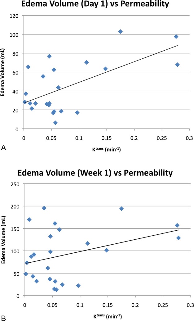
Background: Spontaneous intracerebral hemorrhage (ICH) is associated with blood-brain barrier (BBB) injury, which is a poorly understood factor in ICH pathogenesis, potentially contributing to edema formation and perihematomal tissue injury. We aimed to assess and quantify BBB permeability following human spontaneous ICH using dynamic contrast-enhanced magnetic resonance imaging (DCE MRI). We also investigated whether hematoma size or location affected the amount of BBB leakage.
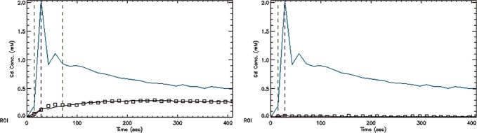
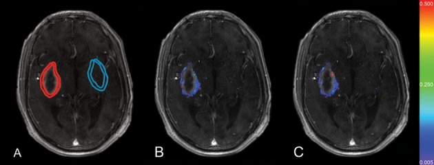
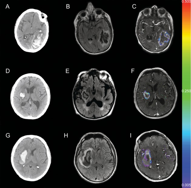
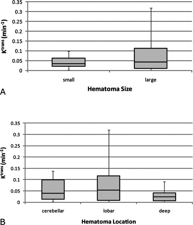
Methods and results: Twenty-five prospectively enrolled patients from the Diagnostic Accuracy of MRI in Spontaneous intracerebral Hemorrhage (DASH) study were examined using DCE MRI at 1 week after symptom onset. Contrast agent dynamics in the brain tissue and general tracer kinetic modeling were used to estimate the forward leakage rate (K(trans)) in regions of interest (ROI) in and surrounding the hematoma and in contralateral mirror-image locations (control ROI). In all patients BBB permeability was significantly increased in the brain tissue immediately adjacent to the hematoma, that is, the hematoma rim, compared to the contralateral mirror ROI (P<0.0001). Large hematomas (>30 mL) had higher K(trans) values than small hematomas (P<0.005). K(trans) values of lobar hemorrhages were significantly higher than the K(trans) values of deep hemorrhages (P<0.005), independent of hematoma volume. Higher K(trans) values were associated with larger edema volumes.
Conclusions: BBB leakage in the brain tissue immediately bordering the hematoma can be measured and quantified by DCE MRI in human ICH. BBB leakage at 1 week is greater in larger hematomas as well as in hematomas in lobar locations and is associated with larger edema volumes.
Keywords: blood–brain barrier; dynamic contrast‐enhanced MRI; intracerebral hemorrhage; magnetic resonance imaging; stroke.
Figures
References
-
- Qureshi AI, Tuhrim S, Broderick JP, Batjer HH, Hondo H, Hanley DF. Spontaneous intracerebral hemorrhage. N Engl J Med. 2001; 344:1450-1460 - PubMed
-
- Morgenstern LB, Hemphill JC, Anderson C, Becker K, Broderick JP, Connolly ES, Jr, Greenberg SM, Huang JN, MacDonald RL, Messe SR, Mitchell PH, Selim M, Tamargo RJ. Guidelines for the management of spontaneous intracerebral hemorrhage: a guideline for healthcare professionals from the American Heart Association/American Stroke Association. Stroke. 2010; 41:2108-2129 - PMC - PubMed
-
- Roger VL, Go AS, Lloyd‐Jones DM, Adams RJ, Berry JD, Brown TM, Carnethon MR, Dai S, de Simone G, Ford ES, Fox CS, Fullerton HJ, Gillespie C, Greenlund KJ, Hailpern SM, Heit JA, Ho PM, Howard VJ, Kissela BM, Kittner SJ, Lackland DT, Lichtman JH, Lisabeth LD, Makuc DM, Marcus GM, Marelli A, Matchar DB, McDermott MM, Meigs JB, Moy CS, Mozaffarian D, Mussolino ME, Nichol G, Paynte NP, Rosamond WD, Sorlie PD, Stafford RS, Turan TN, Turner MB, Wong ND, Wylie‐Rosett J. Heart disease and stroke statistics—2011 update: a report from the American Heart Association. Circulation. 2011; 123:e18-e209 - PMC - PubMed
-
- Flaherty ML, Haverbusch M, Sekar P, Kissela B, Kleindorfer D, Moomaw CJ, Sauerbeck L, Schneider A, Broderick JP, Woo D. Long‐term mortality after intracerebral hemorrhage. Neurology. 2006; 66:1182-1186 - PubMed
-
- Mayer SA, Lignelli A, Fink ME, Kessler DB, Thomas CE, Swarup R, Van Heertum RL. Perilesional blood flow and edema formation in acute intracerebral hemorrhage: a SPECT study. Stroke. 1998; 29:1791-1798 - PubMed
Publication types
MeSH terms
Grants and funding
LinkOut - more resources
Full Text Sources
Other Literature Sources
Medical
