

Suspensor length determines developmental progression of the embryo in Arabidopsis
- PMID: 23709666
- PMCID: PMC3707531
- DOI: 10.1104/pp.113.217166
Suspensor length determines developmental progression of the embryo in Arabidopsis
Abstract
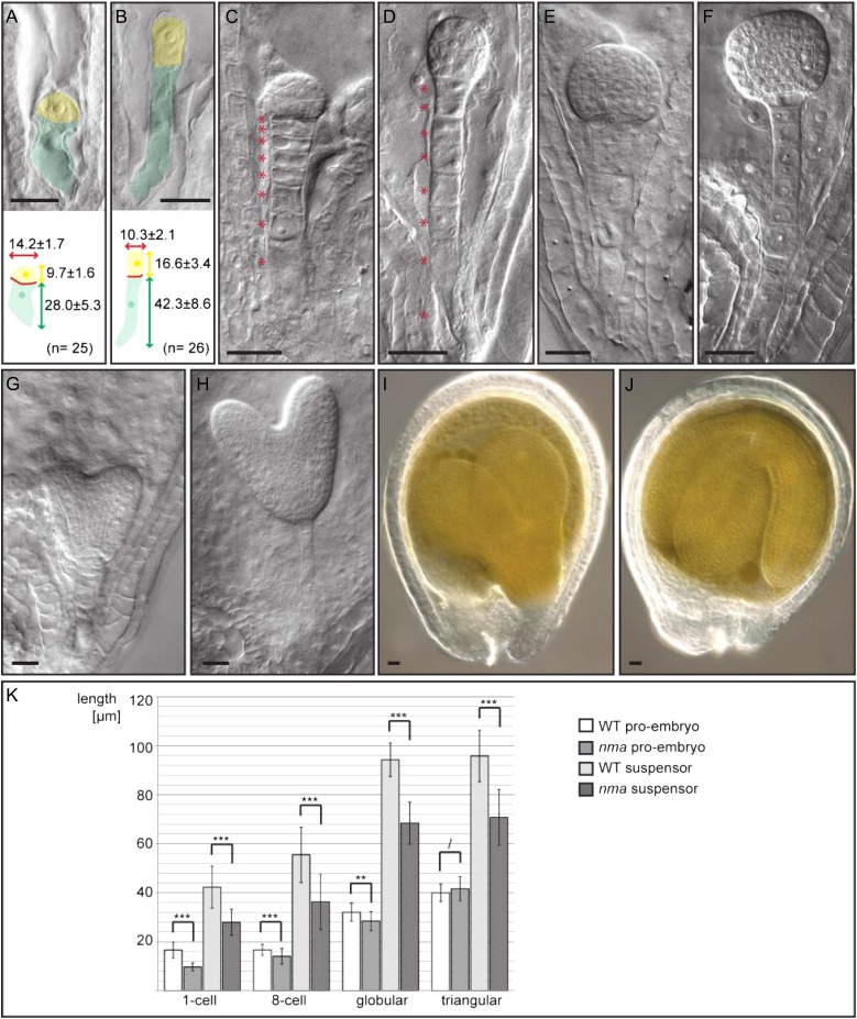
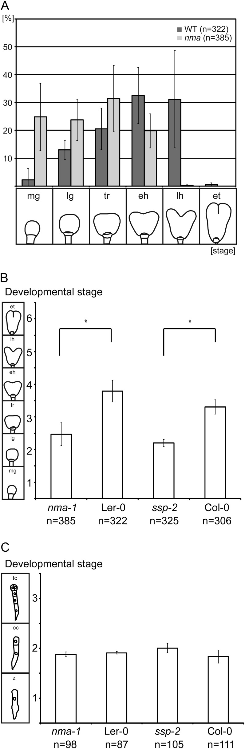
The first structure that differentiates during plant embryogenesis is the extra-embryonic suspensor that positions the embryo in the lumen of the seed. A central role in nutrient transport has been ascribed to the suspensor in species with prominent suspensor structures. Little is known, however, about what impact the size of the rather simple Arabidopsis (Arabidopsis thaliana) suspensor has on embryogenesis. Here, we describe mutations in the predicted exo-polygalacturonase gene NIMNA (NMA) that lead to cell elongation defects in the early embryo and markedly reduced suspensor length. Mutant nma embryos develop slower than wild-type embryos, and we could observe a similar developmental delay in another mutant with shorter suspensors. Interestingly, for both genes, the paternal allele has a stronger influence on the embryonic phenotype. We conclude that the length of the suspensor is crucial for fast developmental progression of the embryo in Arabidopsis.
Figures
Similar articles
-
Direct evidence that suspensor cells have embryogenic potential that is suppressed by the embryo proper during normal embryogenesis.Proc Natl Acad Sci U S A. 2015 Oct 6;112(40):12432-7. doi: 10.1073/pnas.1508651112. Epub 2015 Sep 22. Proc Natl Acad Sci U S A. 2015. PMID: 26396256 Free PMC article.
-
Suspensor-derived somatic embryogenesis in Arabidopsis.Development. 2020 Jul 8;147(13):dev188912. doi: 10.1242/dev.188912. Development. 2020. PMID: 32554529
-
Mutation in the Arabidopisis thaliana DEK1 calpain gene perturbs endosperm and embryo development while over-expression affects organ development globally.Planta. 2005 Jun;221(3):339-51. doi: 10.1007/s00425-004-1448-6. Epub 2005 Jan 13. Planta. 2005. PMID: 15647902
-
The suspensor: not just suspending the embryo.Trends Plant Sci. 2010 Jan;15(1):23-30. doi: 10.1016/j.tplants.2009.11.002. Epub 2009 Dec 4. Trends Plant Sci. 2010. PMID: 19963427 Review.
-
Uncovering the post-embryonic functions of gametophytic- and embryonic-lethal genes.Trends Plant Sci. 2011 Jun;16(6):336-45. doi: 10.1016/j.tplants.2011.02.007. Epub 2011 Mar 17. Trends Plant Sci. 2011. PMID: 21420345 Review.
Cited by
-
Plant Polygalacturonases Involved in Cell Elongation and Separation-The Same but Different?Plants (Basel). 2014 Dec 9;3(4):613-23. doi: 10.3390/plants3040613. Plants (Basel). 2014. PMID: 27135523 Free PMC article. Review.
-
A Profusion of Molecular Scissors for Pectins: Classification, Expression, and Functions of Plant Polygalacturonases.Front Plant Sci. 2018 Aug 14;9:1208. doi: 10.3389/fpls.2018.01208. eCollection 2018. Front Plant Sci. 2018. PMID: 30154820 Free PMC article. Review.
-
Direct evidence that suspensor cells have embryogenic potential that is suppressed by the embryo proper during normal embryogenesis.Proc Natl Acad Sci U S A. 2015 Oct 6;112(40):12432-7. doi: 10.1073/pnas.1508651112. Epub 2015 Sep 22. Proc Natl Acad Sci U S A. 2015. PMID: 26396256 Free PMC article.
-
Plasmogamic Paternal Contributions to Early Zygotic Development in Flowering Plants.Front Plant Sci. 2020 Jun 19;11:871. doi: 10.3389/fpls.2020.00871. eCollection 2020. Front Plant Sci. 2020. PMID: 32636867 Free PMC article. Review.
-
Identification of a gene encoding polygalacturonase expressed specifically in short styles in distylous common buckwheat (Fagopyrum esculentum).Heredity (Edinb). 2019 Oct;123(4):492-502. doi: 10.1038/s41437-019-0227-x. Epub 2019 May 10. Heredity (Edinb). 2019. PMID: 31076649 Free PMC article.
References
-
- Aarssen LW. (2000) Why are most selfers annuals? A new hypothesis for the fitness benefit of selfing. Oikos 89: 606–612
-
- Alonso JM, Stepanova AN, Leisse TJ, Kim CJ, Chen H, Shinn P, Stevenson DK, Zimmerman J, Barajas P, Cheuk R, et al. (2003) Genome-wide insertional mutagenesis of Arabidopsis thaliana. Science 301: 653–657 - PubMed
-
- Barakate A, Martin W, Quigley F, Mache R. (1993) Characterization of a multigene family encoding an exopolygalacturonase in maize. J Mol Biol 229: 797–801 - PubMed
-
- Baud S, Wuillème S, Lemoine R, Kronenberger J, Caboche M, Lepiniec L, Rochat C. (2005) The AtSUC5 sucrose transporter specifically expressed in the endosperm is involved in early seed development in Arabidopsis. Plant J 43: 824–836 - PubMed
-
- Bayer M, Nawy T, Giglione C, Galli M, Meinnel T, Lukowitz W. (2009) Paternal control of embryonic patterning in Arabidopsis thaliana. Science 323: 1485–1488 - PubMed
Publication types
MeSH terms
Substances
LinkOut - more resources
Full Text Sources
Other Literature Sources
Molecular Biology Databases
