. 2013 Apr 9:2013:627240.
doi: 10.1155/2013/627240. Print 2013.

Early neural cell death is an extensive, dynamic process in the embryonic chick and mouse retina

Affiliations

Early neural cell death is an extensive, dynamic process in the embryonic chick and mouse retina

Teresa Chavarría et al. ScientificWorldJournal. .

Abstract

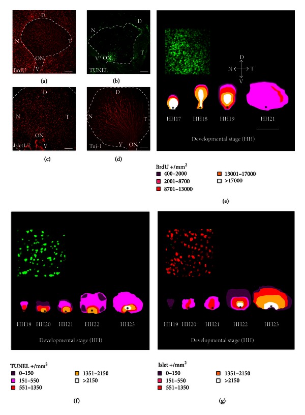
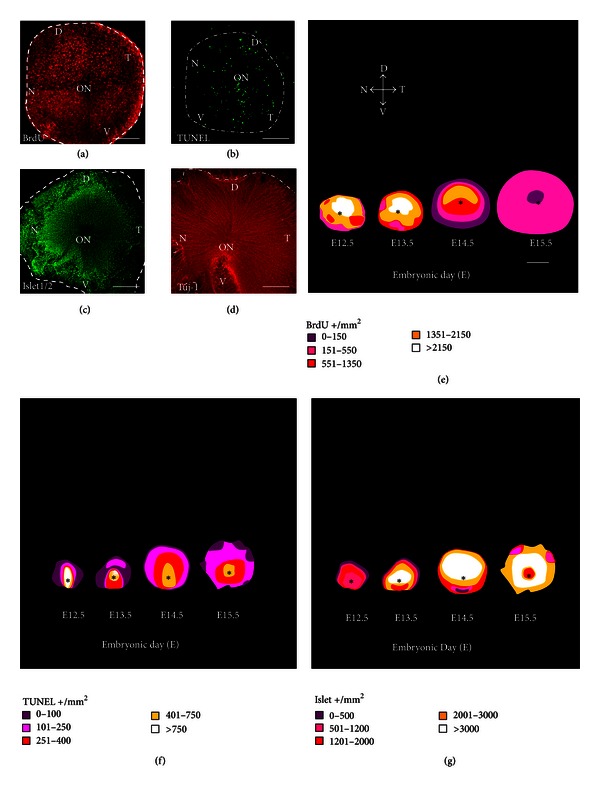
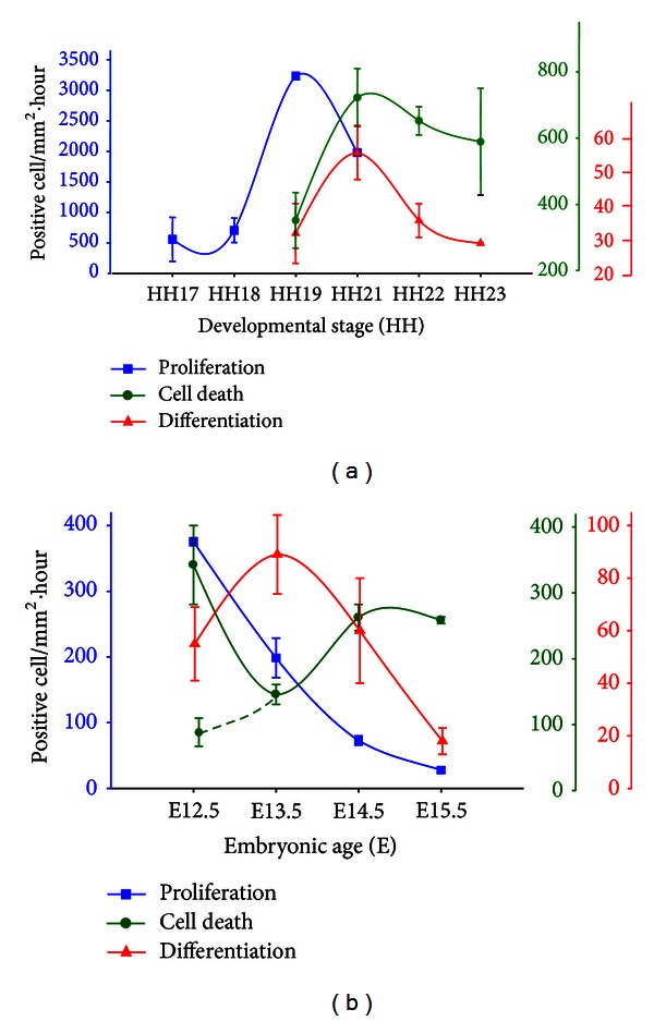
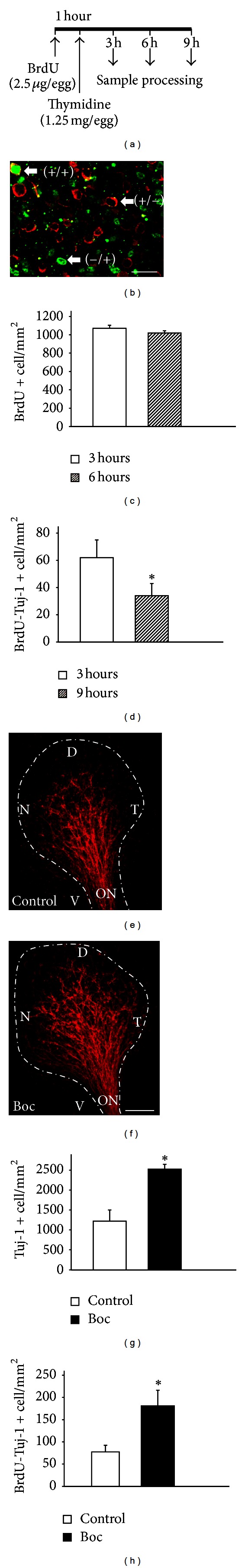
Orchestrated proliferation, differentiation, and cell death contribute to the generation of the complex cytoarchitecture of the central nervous system, including that of the neuroretina. However, few studies have comprehensively compared the spatiotemporal patterns of these 3 processes, or their relative magnitudes. We performed a parallel study in embryonic chick and mouse retinas, focusing on the period during which the first neurons, the retinal ganglion cells (RGCs), are generated. The combination of in vivo BrdU incorporation, immunolabeling of retinal whole mounts for BrdU and for the neuronal markers Islet1/2 and β III-tubulin, and TUNEL allowed for precise cell scoring and determination the spatiotemporal patterns of cell proliferation, differentiation, and death. As predicted, proliferation preceded differentiation. Cell death and differentiation overlapped to a considerable extent, although the magnitude of cell death exceeded that of neuronal differentiation. Precise quantification of the population of recently born RGCs, identified by BrdU and β III-tubulin double labeling, combined with cell death inhibition using a pan-caspase inhibitor, revealed that apoptosis decreased this population by half shortly after birth. Taken together, our findings provide important insight into the relevance of cell death in neurogenesis.


Figures

Figure 1
Figure 1
Overlapping cell processes in the embryonic chick retina. Representative labeling of cell proliferation (a), death (b), and differentiation (c), (d) in retinal whole mounts from chick embryos ((a), HH19; (b), HH20; (c), HH23; (d), HH21). Positive cells were scored in 4–6 retinas from different embryos and represented on isodensity maps of the different processes (e)–(g). The insets show positive cells at higher magnification, as employed for scoring. The main morphological features are labeled as follows. ON and *: optic nerve; D: dorsal; V: ventral; T: temporal; and, N: nasal. Scale bars represent 80 μm (a)–(d), 500 μm (e)–(g), and 40 μm (insets).
Figure 2
Figure 2
Overlapping cell processes in the embryonic mouse retina. Representative labeling of cell proliferation (a), death (b), and differentiation (c), (d) in retinal whole mounts from E13.5 mouse embryos. Positive cells were scored in 3–10 retinas from different embryos and represented on isodensity maps of the different processes (e)–(g). The main morphological features are labeled as follows: ON and *: optic nerve; D: dorsal; V: ventral; T: temporal; N: nasal. Scale bars represent 150 μm (a)–(d) and 300 μm (e)–(g).
Figure 3
Figure 3
Relative magnitudes of cell proliferation, differentiation, and death in embryonic chick and mouse retinas. The results of the experiments described in Figures 1 and 2 were normalized per retinal surface area and hour, as explained in Section 3.2. Graphs represent the relative magnitude of cell proliferation (blue lines: BrdU-positive cells), differentiation (red lines: Islet1/2-positive cells), and death (green lines: TUNEL-positive cells) in chick (a) and mouse (b) embryonic retinas. The alternative slashed line representing cell death in the mouse retina indicates the magnitude of this process after subtracting the TUNEL-positive nuclei associated to the optic fissure, which plays a role in morphogenesis.
Figure 4
Figure 4
Effect of cell death on recently generated retinal ganglion cells. In HH21 chick embryos, in ovo window labeling (a) was used to visualize a defined population of recently generated neurons ((b); +/+; Tuj-1-positive in red; BrdU-positive in green). Proliferating cells ((b); −/+; BrdU-positive in green) were scored at different time points (c) to confirm complete closure of the window. Preexisting neurons were also visualized ((b); +/−; Tuj-1-positive in red). Recently generated neurons were scored at different time points (d). Retinal whole mounts treated with Boc-D-fmk exhibited an expanded neuronal domain as compared with untreated retinas, as visualized by Tuj-1 immunostaining (e) and (f). Total neurons (g) and recently generated neurons (h) were scored in control and Boc-D-fmk-treated embryos. The main morphological features are labeled as follows: ON: optic nerve; D: dorsal; V: ventral; T: temporal; N: nasal. Scale bars represent 20 μm (b) and 80 μm (e), (f). Graphs represent the mean ± SD of at least 3 retinas from different embryos. *P < 0.005.

References

    1. Hamburger V, Levi-Montalcini R. Proliferation, differentiation and degeneration in the spinal ganglia. The Journal of experimental zoology. 1949;111(3):457–501. - PubMed
    1. de la Rosa EJ, de Pablo F. Cell death in early neural development: beyond the neurotrophic theory. Trends in Neurosciences. 2000;23(10):454–458. - PubMed
    1. Yeo W, Gautier J. Early neural cell death: dying to become neurons. Developmental Biology. 2004;274(2):233–244. - PubMed
    1. Buss RR, Sun W, Oppenheim RW. Adaptive roles of programmed cell death during nervous system development. Annual Review of Neuroscience. 2006;29:1–35. - PubMed
    1. McConnell MJ, MacMillan HR, Chun J. Mathematical modeling supports substantial mouse neural progenitor cell death. Neural Development. 2009;4(1, article 28) - PMC - PubMed

Publication types

LinkOut - more resources