. 2013 May;6(5):27-37.

Appearance of New Vemurafenib-associated Melanocytic Nevi on Normal-appearing Skin: Case Series and a Review of Changing or New Pigmented Lesions in Patients with Metastatic Malignant Melanoma After Initiating Treatment with Vemurafenib

Affiliations

Appearance of New Vemurafenib-associated Melanocytic Nevi on Normal-appearing Skin: Case Series and a Review of Changing or New Pigmented Lesions in Patients with Metastatic Malignant Melanoma After Initiating Treatment with Vemurafenib

Philip R Cohen et al. J Clin Aesthet Dermatol. 2013 May.

Abstract

Background: Vemurafenib, a selective BRAF inhibitor that has antineoplastic activity in patients with unresectable or metastatic malignant melanoma whose tumor harbors a BRAF V600E mutation, has multiple drug-associated cutaneous adverse effects.

Purpose: To provide a detailed and comprehensive review of reported changing or new pigmented lesions in oncology patients who have been treated with vemurafenib.

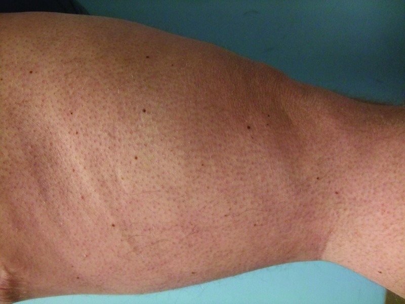
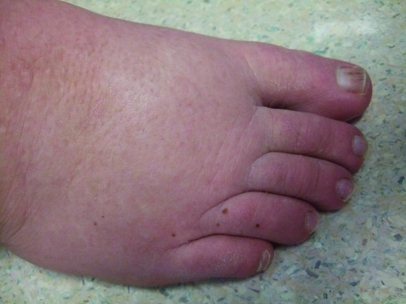
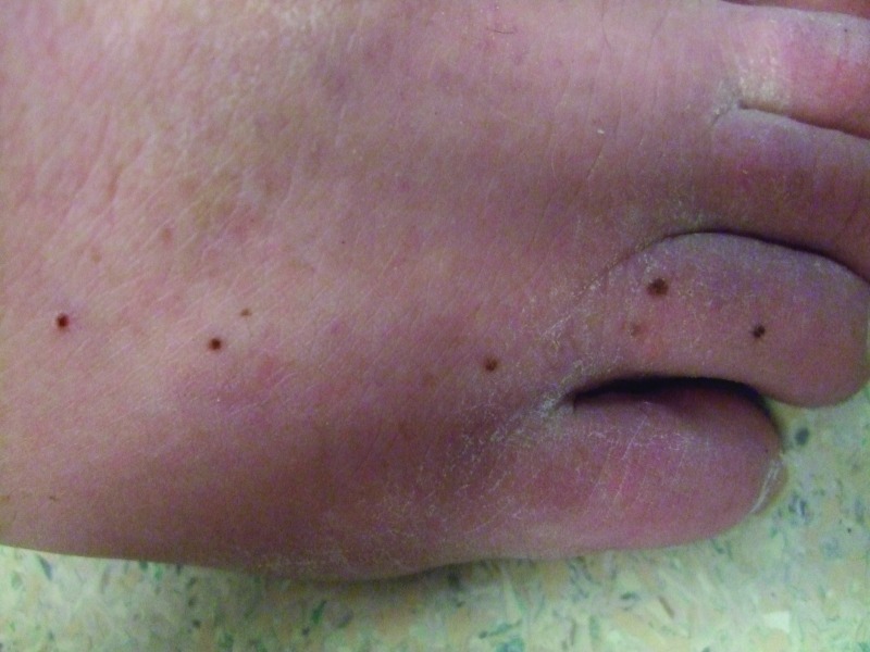
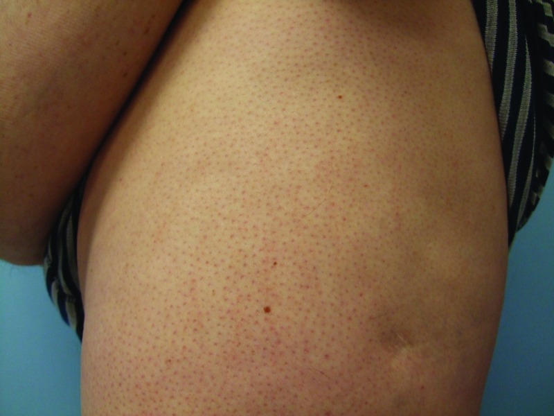
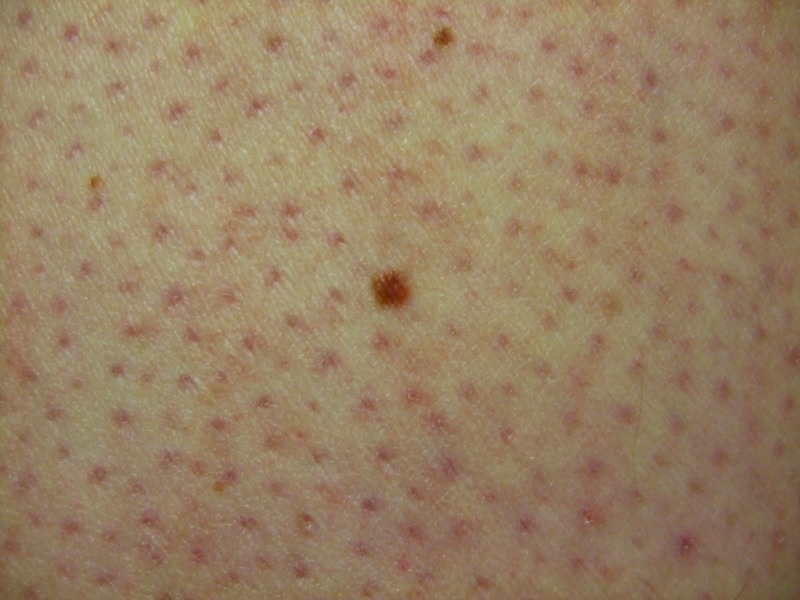
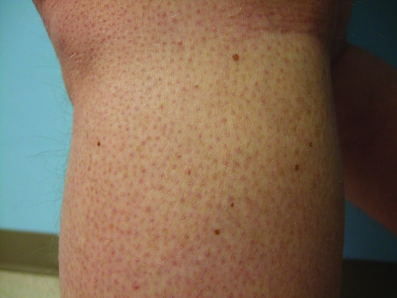
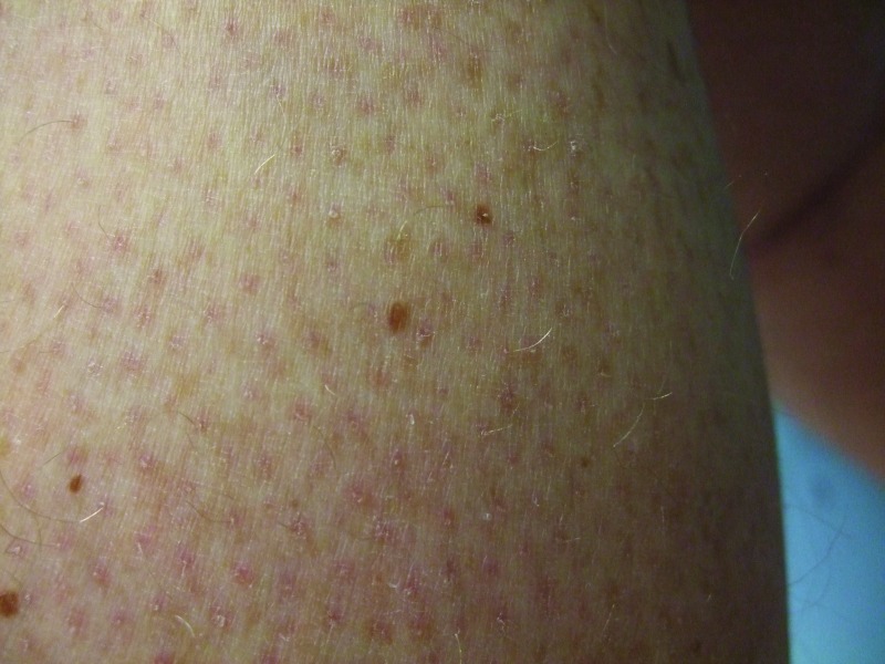
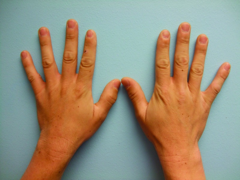
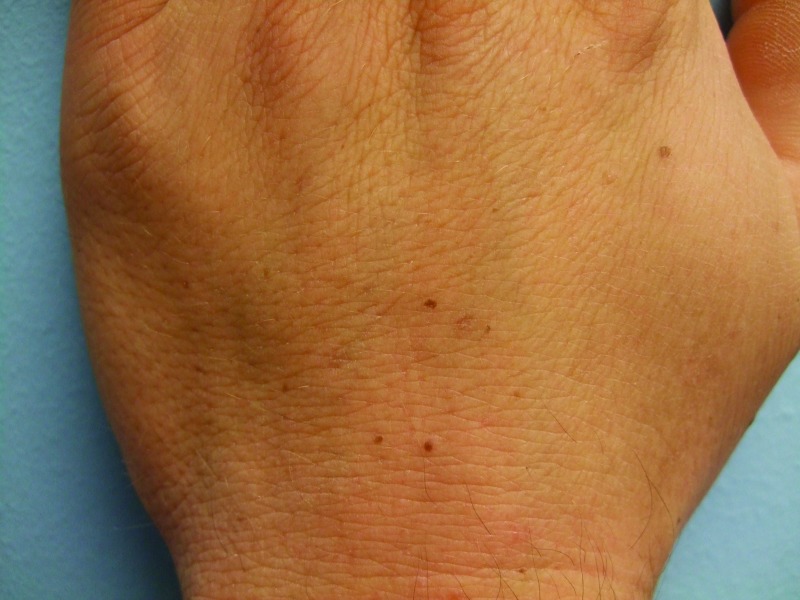
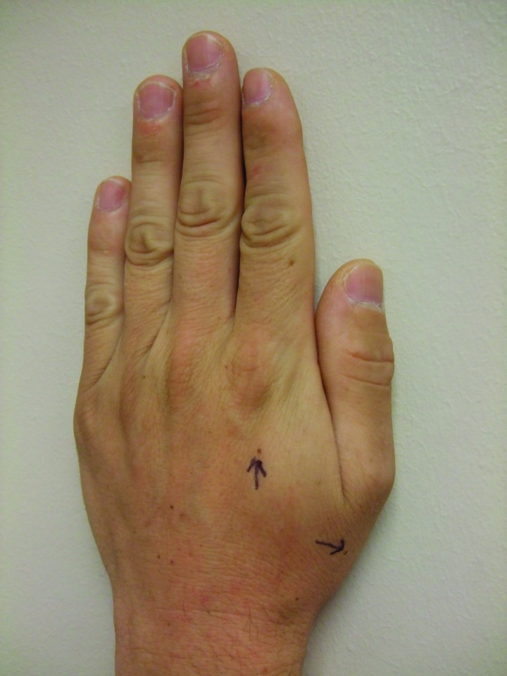
Methods: The new appearance of melanocytic nevi on normal-appearing skin after initiating treatment with vemurafenib is described in two men with metastatic malignant melanoma whose tumors demonstrated a BRAF V600E mutation. Using the PubMed database, an extensive literature search was performed for the following topics: vermurafenib, nevus, nevi, melanoma, pigmented lesion, cutaneous, adverse effect, side effect. The results of the search were used to secure all reports of new or changing pigmented lesions after initiating treatment with vemurafenib.

Results: Vemurafenib is associated with both changes in existing pigmented lesions (including involution, alteration of color and size, and progression to melanoma) and the onset of new melanocytic lesions-nevi (in 5 patients) and primary melanomas (in 2 patients). Visual examination, dermoscopic evaluation, and reflectance confocal microscopy have been used to document the changes in existing or new melanocytic lesions subsequent to initiating treatment with vermurafenib. Histopathology analysis has shown these lesions to usually be either dysplastic nevi or new primary melanomas.

Conclusion: Vemurafenib-treated patients can develop new pigmented lesions (such as nevi) and/or morphological changes in their existing melanocytic lesions (such as involution, increase in size, or alternation of color). In addition, they can develop new primary malignant melanomas that either occur de novo on normal-appearing skin or develop in pre-existing melanocytic lesions. Therefore, total body skin examination should be considered prior to initiating treatment with vemurafenib. Regularly scheduled follow-up skin examinations are also recommended for patients while they are receiving this drug. In addition, for patients who are being treated with vemurafenib, either dermoscopic or photographic or visual modalities should be used to evaluate new or changing pigmented lesions. Also, biopsy for histopathology should be considered for vemurafenib-treated patients who develop new pigmented lesions or whose existing melanocytic lesions have morphological changes in size or color.


Figures

Figures 1A and 1B
Figures 1A and 1B
Posterior-lateral view of left (A) and right (B) proximal legs of a 39-year-old man with a BRAF V600E mutation metastatic malignant melanoma show new pigmented lesions after initiating treatment with vemurafenib.
Figures 1A and 1B
Figures 1A and 1B
Posterior-lateral view of left (A) and right (B) proximal legs of a 39-year-old man with a BRAF V600E mutation metastatic malignant melanoma show new pigmented lesions after initiating treatment with vemurafenib.
Figure 2
Figure 2
The right dorsal foot shows new melanocytic lesions 12 weeks after starting vemurafenib—three nevi on the fourth toe and three nevi on the dorsal foot proximal to his toes.
Figures 3A and 3B
Figures 3A and 3B
Distant (A) and closer (B) views of the lateral left foot show new pigmented lesions on the left heel (1), proximal dorso-lateral foot (2), great toe (1), and fifth toe (1) that appeared three months after vemurafenib therapy was begun.
Figures 3A and 3B
Figures 3A and 3B
Distant (A) and closer (B) views of the lateral left foot show new pigmented lesions on the left heel (1), proximal dorso-lateral foot (2), great toe (1), and fifth toe (1) that appeared three months after vemurafenib therapy was begun.
Figure 4
Figure 4
Another new melanocytic lesion (proximally located) has appeared on the right dorsal foot at follow-up examination four months after beginning vemurafenib treatment; in addition, some of the earlier appearing new pigmented lesions have increased in size.
Figures 5A, 5B, 5C, and 5D
Distant (A and C) and closer (B and D) views of the left lateral thigh (A and B) and left calf (C and D) show the vemurafenib-associated new melanocytic nevi that were biopsied; these were the inferior and larger brown lesion on the thigh (B) and the superior and lateral brown lesion on the calf. Pathology analysis of each pigmented lesions showed a dysplastic nevus characterized by a compound melanocytic nevus with moderate architectural disorder and moderate cytologic atypia. His keratosis pilaris-like eruption is also prominent.
Figures 6A, 6B, and 6C
Distant (A) and closer (B and C) views of the left (B) and right (C) dorsal hands of a 33-year-old man with metastatic malignant melanoma that harbors a BRAF V600E mutation show new melanocytic nevi that appeared two months after initiating vemurafenib treatment—five on the left hand (B) and three on the right hand and third finger.
Figure 7
Figure 7
Newly appearing pigmented lesions—after beginning therapy with vemurafenib—on the left hand that were biopsied (arrows). Pathologic analysis of the lesion proximal to the thumb showed a benign compound melanocytic nevus. Pathologic analysis of the lesion proximal to the index finger showed a dysplastic nevus characterized by a junctional melanocytic nevus with moderate architectural disorder and moderate cytologic atypia; incidentally, spongiosis and focal acantholytic dyskeratosis were also noted.

References

    1. Jordan EJ, Kelly CM. Vemurafenib for the treatment of melanoma. Expert Opin Pharmacother. 2012;13:2533–2543. - PubMed
    1. Ravnan MC, Matalka MS. Vemurafenib in patients with BRAF V600E mutation-positive advanced melanoma. Clin Ther. 2012;34:1474–1486. - PubMed
    1. Fisher R, Larkin J. Vemurafenib: a new treatment for BRAF-V600 mutated advanced melanoma. Cancer Management and Research. 2012;4:243–252. - PMC - PubMed
    1. Flaherty KT. BRAF inhibitors and melanoma. Cancer J. 2011;17:505–511. - PubMed
    1. Sosman JA, Kim KB, Schuchter L, et al. Survival in BRAF V600-mutant advanced melanoma treated with vemurafenib. N Engl J Med. 2012;366:707–714. - PMC - PubMed

LinkOut - more resources