

A rare case of interdigitating dendritic cell sarcoma in the nasal cavity
- PMID: 23710400
- PMCID: PMC3654715
- DOI: 10.1155/2013/913157
A rare case of interdigitating dendritic cell sarcoma in the nasal cavity
Abstract
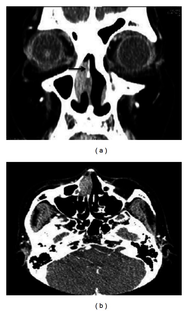
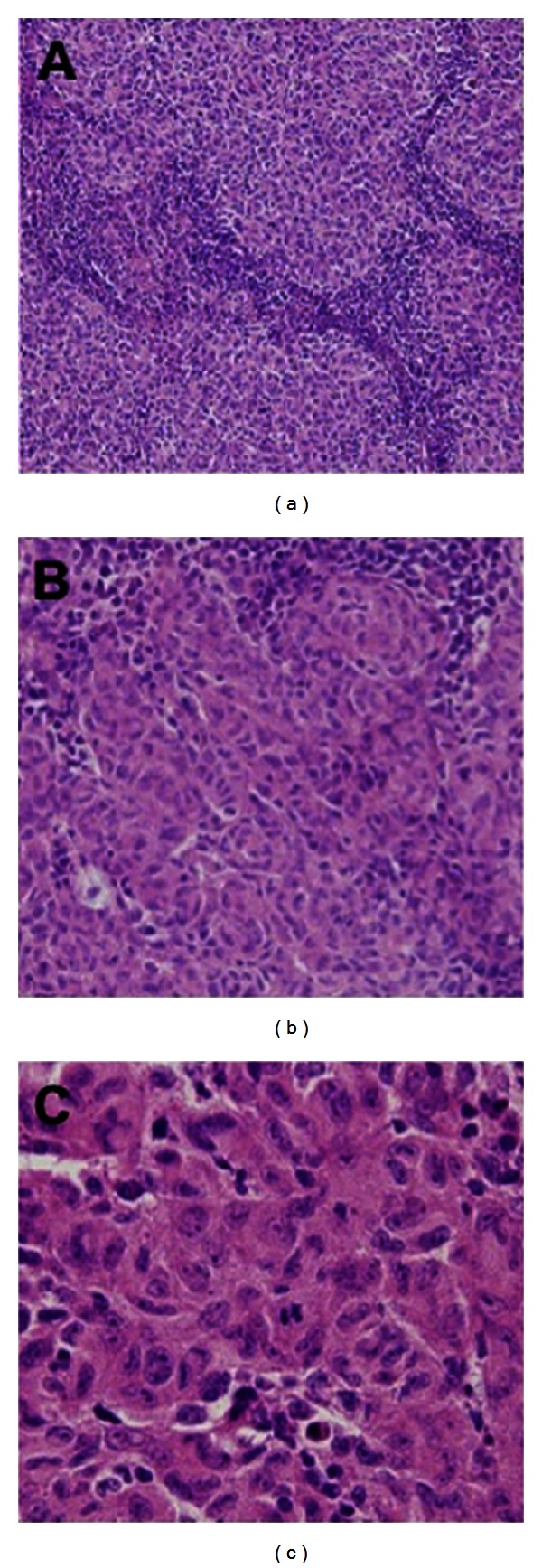
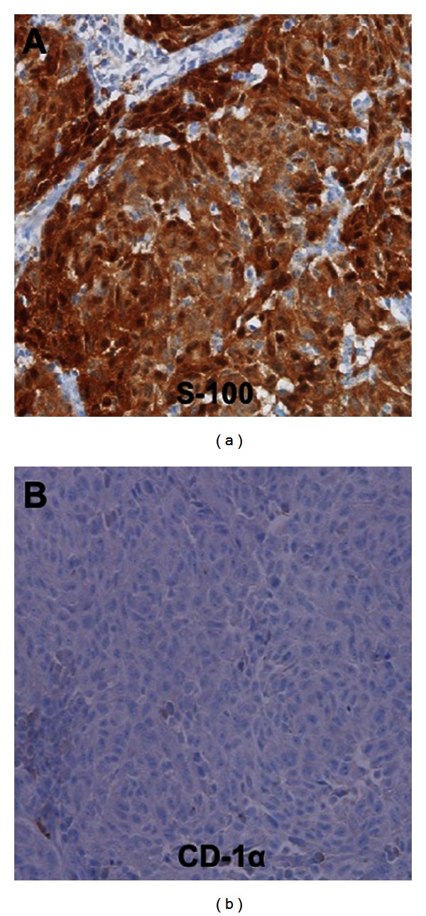
Interdigitating dendritic cell sarcoma (IDCS) is an extremely rare neoplasm that mainly arises from the lymphoid tissues of the immune system. Although this neoplasm typically occurs anywhere along the lymph nodes, it can also be found at extranodal sites, especially in the head and neck. We experienced a rare case of extranodal IDCS in the nasal cavity, a location that has not been previously reported. A 73-year-old woman presented with a polyp-like mass in the nasal cavity and underwent endoscopic sinus surgery. A histologic study confirmed the mass as IDCS by immunohistochemistry with S-100 antibody, and postoperative adjuvant radiotherapy was administered. Although the incidence is extremely rare, this case suggests that extranodal IDCS should be considered in the differential diagnosis of nasal cavity masses.
Figures
Similar articles
-
Head and Neck Extranodal Interdigitating Dendritic Cell Sarcoma: Case Report and Review of the Literature.Head Neck Pathol. 2016 Jun;10(2):145-51. doi: 10.1007/s12105-015-0627-z. Epub 2015 Apr 19. Head Neck Pathol. 2016. PMID: 25893828 Free PMC article. Review.
-
Interdigitating dendritic cell sarcoma presenting in the nasal region.Pathol Res Pract. 2012 Jun 15;208(6):368-71. doi: 10.1016/j.prp.2012.02.004. Epub 2012 May 1. Pathol Res Pract. 2012. PMID: 22554504
-
Extranodal interdigitating dendritic cell sarcoma presenting in the pleura: a case report.J Korean Med Sci. 2011 Feb;26(2):304-7. doi: 10.3346/jkms.2011.26.2.304. Epub 2011 Jan 24. J Korean Med Sci. 2011. PMID: 21286027 Free PMC article.
-
A Case Presentation of a Rare Pelvic Interdigitating Dendritic Cell Sarcoma.Cureus. 2024 Feb 14;16(2):e54220. doi: 10.7759/cureus.54220. eCollection 2024 Feb. Cureus. 2024. PMID: 38371440 Free PMC article.
-
Interdigitating dendritic cell sarcoma and follicular dendritic cell sarcoma: histopathological findings for differential diagnosis.J Clin Exp Hematop. 2013;53(3):179-84. doi: 10.3960/jslrt.53.179. J Clin Exp Hematop. 2013. PMID: 24369219 Review.
Cited by
-
Treatment, outcomes, and demographics in sinonasal sarcoma: a systematic review of the literature.BMC Ear Nose Throat Disord. 2018 Mar 21;18:4. doi: 10.1186/s12901-018-0052-5. eCollection 2018. BMC Ear Nose Throat Disord. 2018. PMID: 29581706 Free PMC article.
-
Head and Neck Extranodal Interdigitating Dendritic Cell Sarcoma: Case Report and Review of the Literature.Head Neck Pathol. 2016 Jun;10(2):145-51. doi: 10.1007/s12105-015-0627-z. Epub 2015 Apr 19. Head Neck Pathol. 2016. PMID: 25893828 Free PMC article. Review.
-
Interdigitating dendritic cell sarcoma presenting in the sigmoid colon mesentery: A case report and literature review.Medicine (Baltimore). 2017 Apr;96(16):e6210. doi: 10.1097/MD.0000000000006210. Medicine (Baltimore). 2017. PMID: 28422825 Free PMC article. Review.
-
Interdigitating dendritic cell sarcoma located in the groin: a case report and literature review.J Int Med Res. 2018 Nov;46(11):4791-4799. doi: 10.1177/0300060518792444. Epub 2018 Sep 17. J Int Med Res. 2018. PMID: 30222020 Free PMC article. Review.
-
Interdigitating dendritic cell sarcoma: analysis of two original extra-nodal cases and review of literature.Virchows Arch. 2022 Jul;481(1):101-110. doi: 10.1007/s00428-022-03320-9. Epub 2022 Apr 9. Virchows Arch. 2022. PMID: 35397699 Review.
References
-
- Perez-Ordonez B, Rosai J. Follicular dendritic cell tumor: review of the entity. Seminars in Diagnostic Pathology. 1998;15(2):144–154. - PubMed
-
- Fonseca R, Yamakawa M, Nakamura S, et al. Follicular dendritic cell sarcoma and interdigitating reticulum cell sarcoma: a review. The American Journal of Hematology. 1998;59(2):161–167. - PubMed
-
- Gaertner EM, Tsokos M, Derringer GA, Neuhauser TS, Arciero C, Andriko JAW. Interdigitating dendritic cell sarcoma: a report of four cases and review of the literature. The American Journal of Clinical Pathology. 2001;115(4):589–597. - PubMed
-
- Andriko JAW, Kaldjian EP, Tsokos M, Abbondanzo SL, Jaffe ES. Reticulum cell neoplasms of lymph nodes: a clinicopathologic study of 11 cases with recognition of a new subtype derived from fibroblastic reticular cells. The American Journal of Surgical Pathology. 1998;22(9):1048–1058. - PubMed
-
- Araújo VC, Martins MT, Salmen FS, Araújo NS. Extranodal follicular dendritic cell sarcoma of the palate. Oral Surgery, Oral Medicine, Oral Pathology, Oral Radiology, and Endodontics. 1999;87(2):209–214. - PubMed
LinkOut - more resources
Full Text Sources
Other Literature Sources
