. 2013 May 28;4(3):57.
doi: 10.1186/scrt209.

Amelioration of motor/sensory dysfunction and spasticity in a rat model of acute lumbar spinal cord injury by human neural stem cell transplantation

Amelioration of motor/sensory dysfunction and spasticity in a rat model of acute lumbar spinal cord injury by human neural stem cell transplantation

Sebastiaan van Gorp et al. Stem Cell Res Ther. .

Abstract

Introduction: Intraspinal grafting of human neural stem cells represents a promising approach to promote recovery of function after spinal trauma. Such a treatment may serve to: I) provide trophic support to improve survival of host neurons; II) improve the structural integrity of the spinal parenchyma by reducing syringomyelia and scarring in trauma-injured regions; and III) provide neuronal populations to potentially form relays with host axons, segmental interneurons, and/or α-motoneurons. Here we characterized the effect of intraspinal grafting of clinical grade human fetal spinal cord-derived neural stem cells (HSSC) on the recovery of neurological function in a rat model of acute lumbar (L3) compression injury.

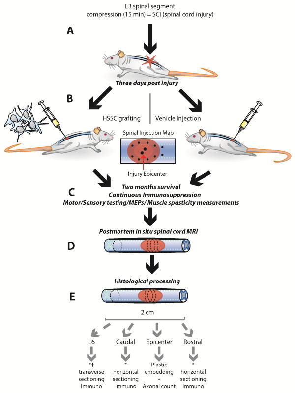
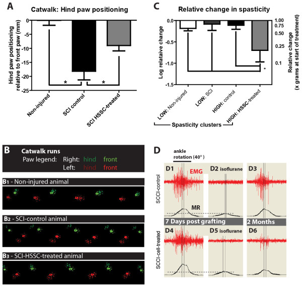
Methods: Three-month-old female Sprague-Dawley rats received L3 spinal compression injury. Three days post-injury, animals were randomized and received intraspinal injections of either HSSC, media-only, or no injections. All animals were immunosuppressed with tacrolimus, mycophenolate mofetil, and methylprednisolone acetate from the day of cell grafting and survived for eight weeks. Motor and sensory dysfunction were periodically assessed using open field locomotion scoring, thermal/tactile pain/escape thresholds and myogenic motor evoked potentials. The presence of spasticity was measured by gastrocnemius muscle resistance and electromyography response during computer-controlled ankle rotation. At the end-point, gait (CatWalk), ladder climbing, and single frame analyses were also assessed. Syrinx size, spinal cord dimensions, and extent of scarring were measured by magnetic resonance imaging. Differentiation and integration of grafted cells in the host tissue were validated with immunofluorescence staining using human-specific antibodies.

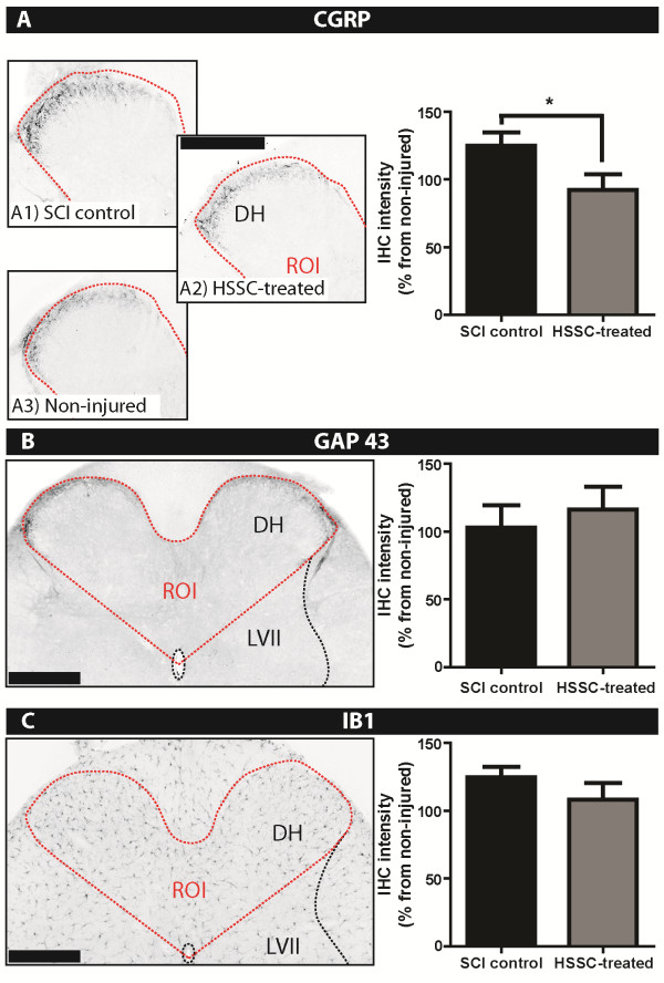
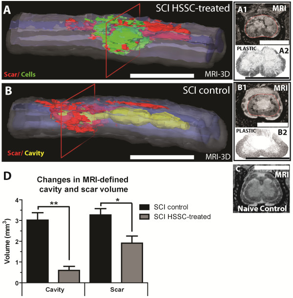
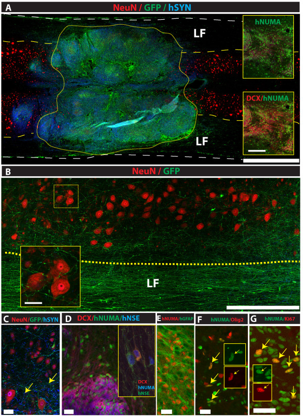
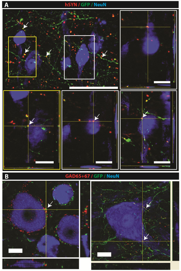
Results: Intraspinal grafting of HSSC led to a progressive and significant improvement in lower extremity paw placement, amelioration of spasticity, and normalization in thermal and tactile pain/escape thresholds at eight weeks post-grafting. No significant differences were detected in other CatWalk parameters, motor evoked potentials, open field locomotor (Basso, Beattie, and Bresnahan locomotion score (BBB)) score or ladder climbing test. Magnetic resonance imaging volume reconstruction and immunofluorescence analysis of grafted cell survival showed near complete injury-cavity-filling by grafted cells and development of putative GABA-ergic synapses between grafted and host neurons.

Conclusions: Peri-acute intraspinal grafting of HSSC can represent an effective therapy which ameliorates motor and sensory deficits after traumatic spinal cord injury.


Figures

Figure 1
Figure 1
Schematic diagram of experimental design. A: To induce spinal cord injury, a 35 g circular rod was placed on the exposed L3 spinal segment and the spinal cord compressed in the dorso-ventral direction for 15 minutes. B: Three days after injury, the animals were randomly assigned to experimental groups and received a spinal graft of HSSC or media only. A total of 12 injections were performed targeting the injury epicenter and adjacent areas (see Spinal Injection Map). C: After spinal injections, the animals survived for two months while being continuously immunosuppressed and periodically tested for recovery of motor/sensory functions, changes in motor evoked potentials (MEPs) and gastrocnemius muscle spasticity response evoked by computer-controlled ankle rotation. D: At two months after treatment, animals were perfusion fixed with 4% PFA and spinal cord MRI-imaged in situ before histological processing. E: After MRI imaging, spinal cords were dissected from the spinal column and spinal blocks prepared for plastic embedding (injury epicenter region) or cryostat sectioning and used for immunofluorescence staining (the regions just above and below the injury epicenter). HSSC, human fetal spinal cord-derived neural stem cells; MRI, magnetic resonance imaging; PFA, paraformaldehyde.
Figure 2
Figure 2
Significant decrease in the dorsal horn CGRP immunoreactivity caudal to the injury epicenter in SCI-HSSC-treated versus SCI-control animals. CGRP- (A), GAP-43- (B), and Iba1- (C) immunoreactivity in the dorsal horns (DH) caudal of the injury epicenter at two months after L3 SCI. The region of interest (ROI) was defined as outlined in B and C (left panels, red dotted line). A: The quantitative densitometry analysis of CGRP-immunostained images in the dorsal horns of SCI-HSSC-treated animals (A2) showed significantly decreased CGRP expression when compared to SCI-control animals (A1). B, C: The dorsal horn GAP-43 or Iba1 immunoreactivity was not significantly different between experimental groups. (A - C: data expressed as mean ± SEM; student t-tests). (Scale Bars: A - C: 500 μm). CRGP, calcitonin gene-related peptide; GAP-13, growth associated protein 43; HSSC, human fetal spinal cord-derived neural stem cells; Iba1, ionized calcium binding adaptor molecule 1; SCI, spinal cord injury.
Figure 3
Figure 3
Improvement in hind paw positioning and muscle spasticity in SCI animals grafted with HSSC. A: CatWalk gait analysis of hind paw positioning at two months after treatment. In comparison to SCI control animals, a significant improvement was seen in HSSC-grafted animals. B1-B3: An example of paw step images taken from the CatWalk software in naïve (B1), SCI-control (B2) and SCI-HSSC-treated animals (B3). Note a large paw footprint overlap between the front and hind paws in naïve animals (B1) but a substantial dissociation in footprint overlap in SCI controls (B2). An improvement in paw placement in SCI-HSSC-treated animals can be seen (B3). C: Statistical analysis showed significant suppression of spasticity response (expressed as a muscle resistance ratio: values at two months versus seven days post injury in ‘HIGH spasticity’ HSSC-treated animals if compared to ‘HIGH spasticity’ controls). D: To identify the presence of muscle spasticity in fully awake animals, the hind-paw ankle is rotated 40° at a velocity of 80°/second. Spasticity is identified by exacerbated EMG activity measured in the gastrocnemius muscle and corresponding increase in muscle resistance. In control SCI animals with developed spasticity (that is, ‘high spasticity’/HIGH group), no change in spasticity response if compared to seven days post-vehicle injection was seen at two months (compare D1 to D3). In contrast to SCI control animals, a decrease in spasticity response was seen in SCI-HSSC-treated animals at two months after cell injections (compare D4 to D6). To identify mechanical resistance, animals are anesthetized with isoflurane at the end of the recording session and the contribution of mechanical resistance (which is, isoflurane non-sensitive) is calculated. (D2, D5: data expressed as mean ± SEM; one-way ANOVAs). ANOVA, analysis of variance; EMG, electromyography; HSSC, human fetal spinal cord-derived neural stem cells; SCI, spinal cord injury; SEM, standard error of the mean.
Figure 4
Figure 4
Amelioration of hypoesthesia in SCI-HSSC-grafted animals. Baseline and biweekly assessments of perceptive thresholds for (A) mechanical and (B) thermal stimuli, applied below the level of injury, showed a trend towards progressive recovery in SCI-HSSC-grafted animals. C: When expressed as percentages of the maximal possible effect for mechanical and thermal perceptive thresholds improvements, SCI-HSSC-treated animals showed significant improvements in sensory function for both mechanical and thermal components. (A-C: data expressed as mean ± SEM; A-B: repeated measures ANOVAs; C: Student t-tests). ANOVA, analysis of variance; HSSC, human fetal spinal cord-derived neural stem cells; SCI, spinal cord injury; SEM, standard error of the mean.
Figure 5
Figure 5
Effective cavity-filling effect by transplanted cells in SCI HSSC-injected animals. At the end of the two-month post-treatment survival, animals were perfusion fixed with 4% PFA, the spinal column dissected and MRI-imaged in situ before spinal cord dissection for further histological processing. A, B: Three-dimensional MRI images of spinal cord segments in animals with previous traumatic injury and treated with spinal HSSC (A) or media (B) injections. Note the near complete injected-cells cavity-filling effect in HSSC-treated animals. A1, A2, B1, B2: To validate the presence of grafted cells or cavitation at the epicenter of injury, the same region was histologically processed, semi-thin plastic sections prepared and compared to the corresponding MRI image (compare A1 to A2 and B1 to B2). C: Two-dimensional MRI image taken from a naïve-non-injured animal. D: Quantification of the cavity and scar volume from serial MRI images showed significantly decreased cavity and scar volumes in SCI-HSSC-injected animals if compared to media-injected SCI controls. (D: data expressed as mean ± SEM; Student t-tests), (Scale Bars: A, B: 5 mm; A1, A2, B1, B2, C: 3 mm). HSSC, human fetal spinal cord-derived neural stem cells; MRI, magnetic resonance imaging; PFA, paraformaldehyde; SCI, spinal cord injury; SEM, standard error of the mean.
Figure 6
Figure 6
Survival, differentiation and extensive axonal outgrowth from spinally grafted HSSC. A: Grafted GFP+ or hNUMA+ cells can be seen almost completely filling the lesion cavity at eight weeks after grafting (yellow dotted area; inserts). B: Detail from ‘A’ depicting a dense GFP+ neurite network in the lateral funiculus (LF) and with numerous axons projecting towards α-motoneurons and interneurons in the gray matter (insert). C: In areas with a dense GFP+ axodendritic network, clear hSYN immunoreactivity associated with GFP+ processes can be detected (yellow arrows). D: The majority of grafted hNUMA+ cells showed development of the neuronal hNSE/DCX+ phenotype. E, F: A subpopulation of grafted hNUMA+ cells showed the astrocyte (hGFAP+) and oligodendrocyte (Olig 2) phenotype (F; yellow arrows). G: Using mitotic marker Ki67, regularly distributed hNUMA/Ki67+ grafted cells were identified (yellow arrows). (Scale Bars: A: 1.5 mm (inserts: 200 μm); B: 600 μm (insert: 75 μm); C: 60 μm; D: 20 μm; E-G: 10 μm). HSSC, human fetal spinal cord-derived neural stem cells; SCI, spinal cord injury.
Figure 7
Figure 7
Development of putative GABA-ergic synaptic contact between HSSC and the host neurons. A: Confocal analysis of hSYN/GFP/NeuN-stained sections shows numerous hSYN punctata associated with GFP+ processes derived from grafted cells. Some of the hSYN/GFP+ terminals were found to be in the vicinity of the host interneurons or α-motoneurons (A; inserts; white arrows). B: Triple staining with GAD65/67/GFP/NeuN antibody showed numerous double-stained GAD65/67/GFP+ terminals residing on or in the close vicinity of lumbar α-motoneurons (white arrows). (Scale Bars: A: 150 μm (inserts: 30 μm); B: 20 μm). HSSC, human fetal spinal cord-derived neural stem cells.

Comment in

Similar articles

Cited by

References

    1. Hagg T, Oudega M. Degenerative and spontaneous regenerative processes after spinal cord injury. J Neurotrauma. 2006;23:264–280. - PubMed
    1. Jones CF, Cripton PA, Kwon BK. Gross morphological changes of the spinal cord immediately after surgical decompression in a large animal model of traumatic spinal cord injury. Spine. 2012;37:E890–E899. doi: 10.1097/BRS.0b013e3182553d1d. - DOI - PubMed
    1. Oudega M. Molecular and cellular mechanisms underlying the role of blood vessels in spinal cord injury and repair. Cell Tissue Res. 2012;349:269–288. doi: 10.1007/s00441-012-1440-6. - DOI - PubMed
    1. Bracken MB. Steroids for acute spinal cord injury. Cochrane Database Syst Rev. 2012;1 CD001046. - PMC - PubMed
    1. Fleming JC, Norenberg MD, Ramsay DA, Dekaban GA, Marcillo AE, Saenz AD, Pasquale-Styles M, Dietrich WD, Weaver LC. The cellular inflammatory response in human spinal cords after injury. Brain. 2006;129:3249–3269. doi: 10.1093/brain/awl296. - DOI - PubMed

Publication types