Comparative Study
. 2013 May 28:11:139.
doi: 10.1186/1741-7015-11-139.

Tissue-derived mesenchymal stromal cells used as vehicles for anti-tumor therapy exert different in vivo effects on migration capacity and tumor growth

Affiliations
Comparative Study

Tissue-derived mesenchymal stromal cells used as vehicles for anti-tumor therapy exert different in vivo effects on migration capacity and tumor growth

Carolina Belmar-Lopez et al. BMC Med. .

Abstract

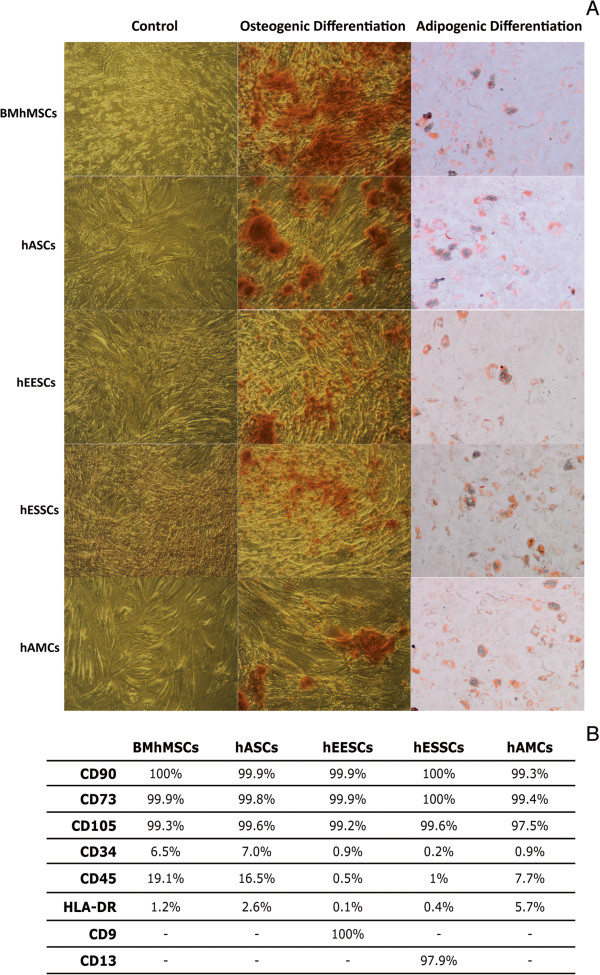
Background: Mesenchymal stem cells (MSCs) have been promoted as an attractive option to use as cellular delivery vehicles to carry anti-tumor agents, owing to their ability to home into tumor sites and secrete cytokines. Multiple isolated populations have been described as MSCs, but despite extensive in vitro characterization, little is known about their in vivo behavior.The aim of this study was to investigate the efficacy and efficiency of different MSC lineages derived from five different sources (bone marrow, adipose tissue, epithelial endometrium, stroma endometrium, and amniotic membrane), in order to assess their adequacy for cell-based anti-tumor therapies. Our study shows the crucial importance of understanding the interaction between MSCs and tumor cells, and provides both information and a methodological approach, which could be used to develop safer and more accurate targeted therapeutic applications.

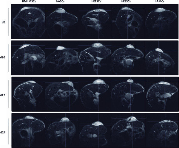
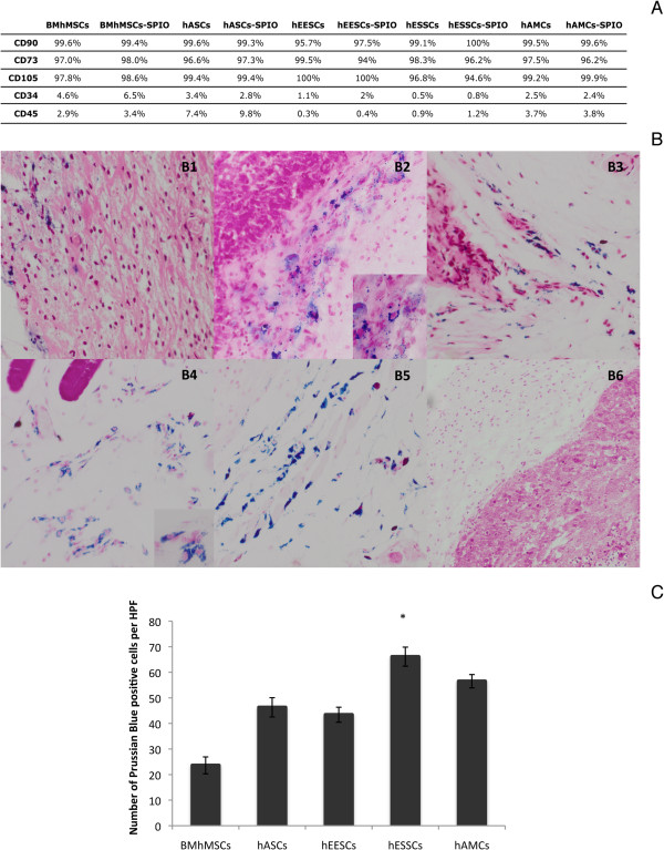
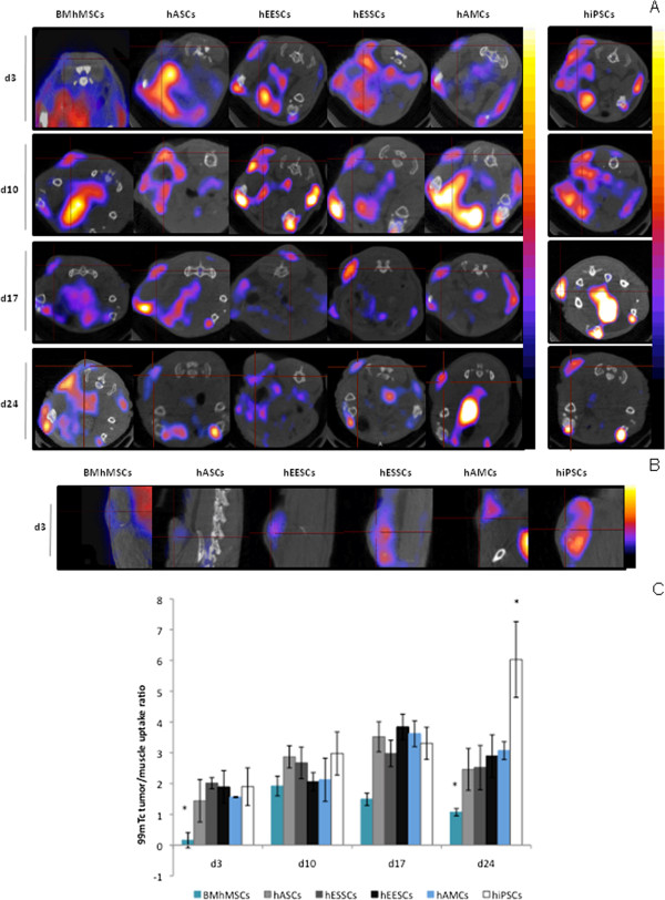
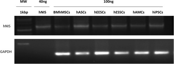
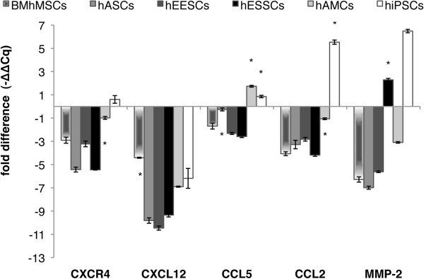
Methods: We first measured the in vivo migration capacity and effect on tumor growth of the different MSCs using two imaging techniques: (i) single-photon emission computed tomography combined with computed tomography (SPECT-CT), using the human sodium iodine symporter gene (hNIS) and (ii) magnetic resonance imaging using superparamagnetic iron oxide. We then sought correlations between these parameters and expression of pluripotency-related or migration-related genes.

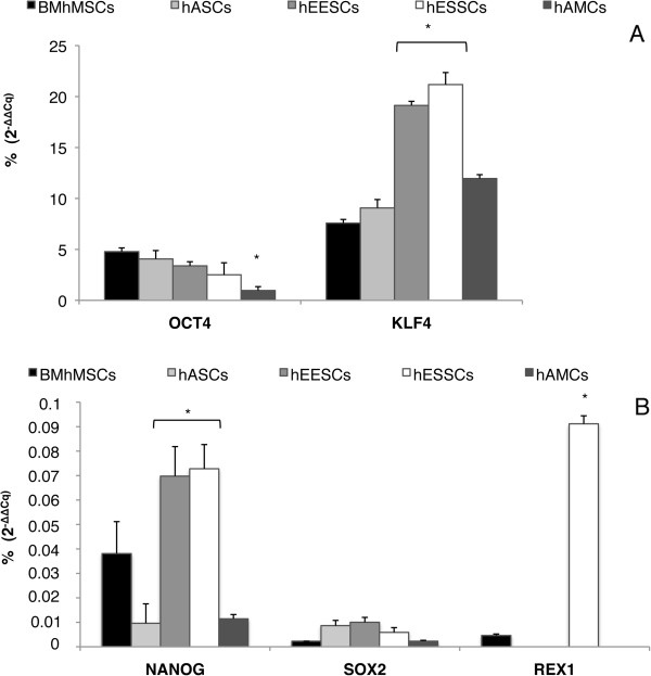
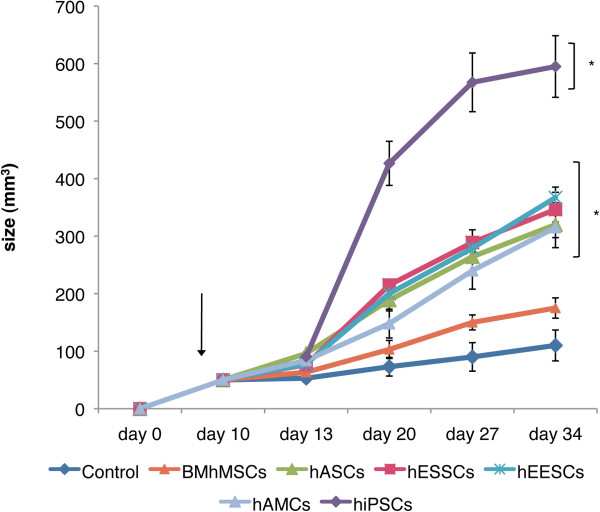
Results: Our results show that migration of human bone marrow-derived MSCs was significantly reduced and slower than that obtained with the other MSCs assayed and also with human induced pluripotent stem cells (hiPSCs). The qPCR data clearly show that MSCs and hiPSCs exert a very different pluripotency pattern, which correlates with the differences observed in their engraftment capacity and with their effects on tumor growth.

Conclusion: This study reveals differences in MSC recruitment/migration toward the tumor site and the corresponding effects on tumor growth. Three observations stand out: 1) tracking of the stem cell is essential to check the safety and efficacy of cell therapies; 2) the MSC lineage to be used in the cell therapy needs to be carefully chosen to balance efficacy and safety for a particular tumor type; and 3) different pluripotency and mobility patterns can be linked to the engraftment capacity of the MSCs, and should be checked as part of the clinical characterization of the lineage.


Figures

Figure 1
Figure 1
Characterization of MSCs. (A) Multi-lineage differentiation potential in vitro to give rise to osteoblasts and adipocytes. Morphologic changes and expression of specific markers for each tissue are shown. (B) Specific surface markers expression (%) detected by flow cytometry.
Figure 2
Figure 2
In vivo xenograft imaging of mice using a magnetic resonance labeling (MRI) scanner. Scans were performed at 3, 10, 17, and 24 days after intravenous injection of super-paramagnetic iron oxide (SPIO)-labeled mesenchymal stem cells (MSCs). Tumors were visible as lighter areas in transverse sections. The recruitment of SPIO-labeled MSCs to tumors resulted in decrease of signal intensity (SI) and the visualization of darker areas in tumor sites. The same animals are represented over the entire period.
Figure 3
Figure 3
Super-paramagnetic iron oxide (SPIO) labeling analysis. (A) Phenotypic analysis after SPIO labeling. Prussian blue staining in tumor sections obtained from animals scanned by MRI at day 24. (B) Images show intense blue clusters in tumor sections. (B1) SPIO-labeled bone marrow-derived human mesenchymal stem cells (BM-hMSCs); (B2) SPIO-labeled human adipose-derived stem cell (hASCs); (B3) SPIO-labeled human epithelial endometrium-derived stem cell (hEESCs); (B4) SPIO-labeled human stromal endometrium-derived stem cells (hESSCs); (B5) SPIO-labeled human amniotic membrane mesenchymal stem cells (hAMCs; (B6) control cells. Original magnification: (B1-B5) ×20; (B6) ×10. (C) number of Prussian blue-stained positive cells per high-power field (HPF) in tumor sections.
Figure 4
Figure 4
In vivo xenograft imaging of mice using a nano-single-photon emission computed tomography/X-ray computed tomography (nano-SPECT-CT) scanner. Scans were performed at 3, 10, 17, and 24 days after intravenous injection of human sodium iodine symporter (hNIS)-labeled mesenchymal stem cells (MSCs) and human induced pluripotent stem cells (hiPSCs). An intravenous dose of 18.5 MBq of 99mTc was administered before the acquisitions. The same animals are represented over the entire period. hNIS expression is indicated by red crosses in transverse sections. Scans were performed after intravenous injection of hNIS-labeled MSCs or hiPSCs. (A) Images at days 3, 10, 17 and 24; (B) images at day 3; (C): 99mTc tumor/muscle uptake ratio by hNIS-labeled MSCs or hiPSCs from days 3 to 24.
Figure 5
Figure 5
Detection of human sodium iodine symporter (hNIS) expression by reverse transcription (RT)-PCR. RNA was extracted from tumors of animals scanned by single-photon emission computed tomography/X-ray computed tomography (SPECT-CT) at day 24.
Figure 6
Figure 6
Pluripotency-related gene expression by quantitative (q)PCR. Gene expression levels were normalized against 18S rRNA. The ratio of the relative expression for each gene to 18S was calculated by using the 2-ΔΔCq formula. hiPSC data were used as a calibrator for the results presented (100%). (A) OCT4 and KLF4; (B) NANOG, SOX2 and REX1.
Figure 7
Figure 7
Effect on xenograft tumor growth. When tumors reached a size of 50 mm3, the animals received an intravenous injection of mesenchymal stem cells (MSCs; arrow). The size of the tumors was measured until the end of the experiments (34 days after the HeLa injection and 24 days after the MSC injection).
Figure 8
Figure 8
Migration-related gene expression by quantitative (PCR). CXCR4, CXCL12, CCL5, CCL2, and MMP-2 expression in tumors (control group) was compared with expression in tumors consisting of HeLa cells and mesenchymal stem cells (MSCs) or of HeLa cells and human induced pluripotent stem cells (hiPSCs) (analyzed 24 days after stem cell injection). The ratio of the fold difference expression for each gene to 18S was calculated by using the -ΔΔCq formula. Expression obtained from the control group was used as a calibrator.

References

    1. Kansas GS. Selectins and their ligands: current concepts and controversies. Blood. 1996;88:3259–3287. - PubMed
    1. Rüster B, Göttig S, Ludwig RJ, Bistrian R, Müller S, Seifried E, Gille J, Henschler R. Mesenchymal stem cells display coordinated rolling and adhesion behavior on endothelial cells. Blood. 2006;108:3938–3944. - PubMed
    1. Schweitzer KM, Dräger AM, van der Valk P, Thijsen SF, Zevenbergen A, Theijsmeijer AP, van der Schoot CE, Langenhuijsen MM. Constitutive expression of E-selectin and vascular cell adhesion molecule-1 on endothelial cells of hematopoietic tissues. Am J Pathol. 1996;148:165–175. - PMC - PubMed
    1. De Miguel MP, Fuentes-Julian S, Blazquez-Martinez A, Pascual CY, Aller MA, Arias J, Arnalich-Montiel F. Immunosuppressive properties of mesenchymal stem cells: advances and applications. Curr Mol Med. 2012;12:574–591. - PubMed
    1. Salem HK, Thiemermann C. Mesenchymal stromal cells: current understanding and clinical status. Stem Cells. 2010;28:585–596. - PMC - PubMed

Publication types

MeSH terms