. 2013 Oct;174(1):38-44.
doi: 10.1111/cei.12146.

Melanocyte-specific CD8+ T cells are associated with epidermal depigmentation in a novel mouse model of vitiligo

Affiliations

Melanocyte-specific CD8+ T cells are associated with epidermal depigmentation in a novel mouse model of vitiligo

S You et al. Clin Exp Immunol. 2013 Oct.

Abstract

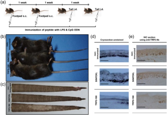
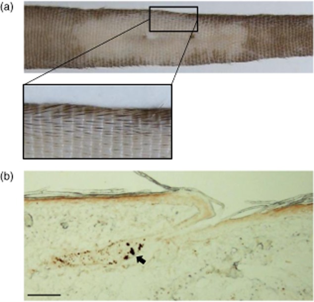
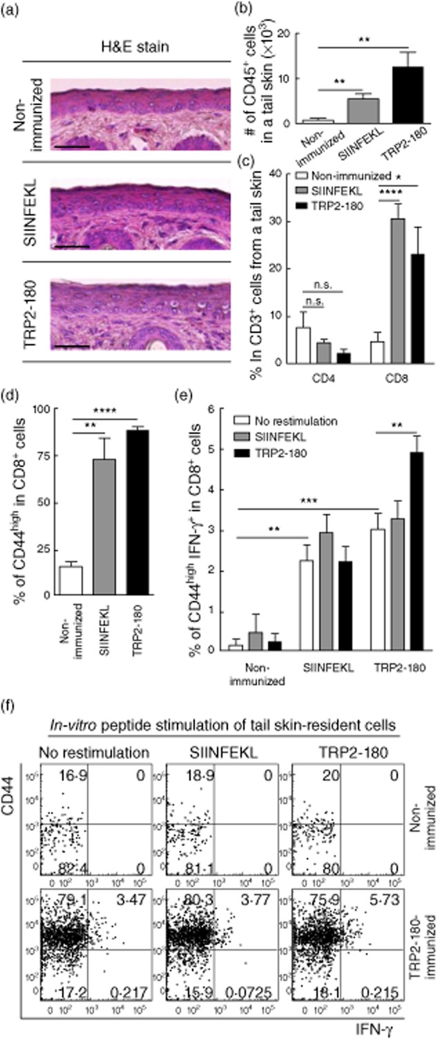
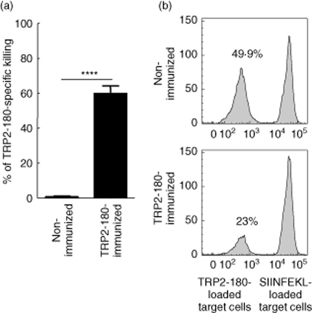
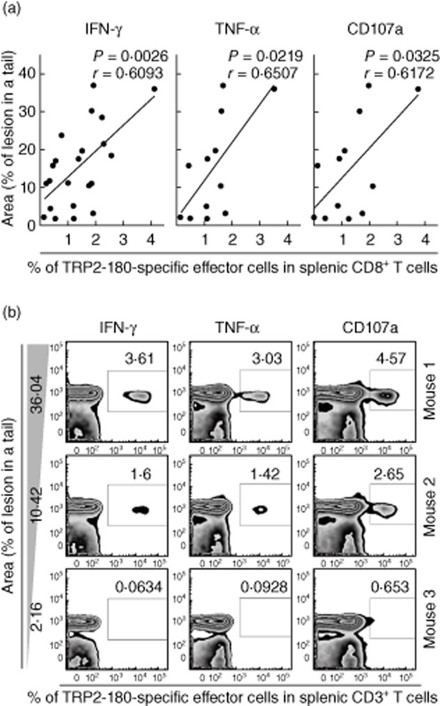
In the present study, we established a novel murine model of vitiligo by sequential prime/boost immunizations into the hind footpad and tail dermis with tyrosinase-related protein 2 (TRP2)-180 (SVYDFFVWL) peptide, lipopolysaccharides and cytosine-phosphate-guanosine (CpG) oligodeoxynucleotides. Immunized mice developed epidermal depigmentation in the tail skin without hair depigmentation, thereby differentiating this approach from established models of vitiligo. Following intradermal tail immunization, activated CD8(+) interferon (IFN)-γ(+) T cells were recruited locally to the tail skin. In-vivo cytotoxicity assays demonstrated specific lysis of TRP2-180-presenting cells in immunized mice. Furthermore, the extent of skin depigmentation correlated with the frequency of TRP2-180-specific splenic CD8(+) T cells, as determined by IFN-γ and tumour necrosis factor (TNF)-α production, and cytotoxic degranulation evidenced by CD107a staining. These findings suggest a correlation between the presence of TRP2-180-specific CD8(+) effector T cells and the development of depigmented skin lesions in our vitiligo model. This new model of vitiligo, characterized by skin depigmentation without hair depigmentation, is more similar to human disease than previous murine models. Therefore, this model is well suited to future studies on the pathogenesis of vitiligo and the development of novel therapeutics for vitiligo.

Keywords: autoreactive CD8+ T cell; epidermal depigmentation; mouse model; vitiligo.


Figures

Fig. 1
Fig. 1
Peptide immunization schedule and development of vitiligo in mice. (a) Peptide immunization schedule. C57BL/6 mice were immunized subcutaneously twice at a 1-week interval in the footpad with tyrosinase-related protein (TRP)2-180 (50 μg), lipopolysacchride (LPS) (5 μg) and cytosine–phosphate–guanosine (CpG) oligodeoxynucleotides (ODN) (5 μg). For boost immunizations, TRP2-180 (50 μg) with the same adjuvants was injected twice intradermally at a 1-week interval into the tail dermis. Control mice were either not immunized or were immunized with SIINFEKL (50 μg) instead of TRP2-180. s.c.: subcutaneous; i.d.: intradermal. (b,c) Gross examination of the tail skin. Five weeks after the last immunization, TRP2-180-immunized mice developed depigmented skin lesions in the tail, but control mice did not (b). Flayed tail skins from these mice are shown (c). (d,e) Histological examination of the tail skin. Depigmented skin lesions of TRP2-180-immunized mice revealed the absence of melanin in unstained cryosections (30 μm) (d) and the absence of melanocytes in immunohistochemistry sections (5 μm) using anti-TRP2 antibodies (e). Scale bars = 200 μm.
Fig. 2
Fig. 2
Hair colour and hair follicle melanocytes in depigmented skin lesions. (a) Hair colour in the depigmented skin lesion. In C57BL/6 mice, a black hair stripe is present on the dorsal part of tail. The photograph shows a representative tail with vitiligo 4 months after depigmentation developed. (b) Immunohistochemistry using anti-tyrosinase-related protein (TRP)2 antibodies shows intact hair follicle melanocytes, whereas epidermal melanocytes were not detected. Arrow indicates melanocytes in the hair follicle. Scale bar = 200 μm.
Fig. 3
Fig. 3
Recruitment and cytokine production of CD8+ T cells in the tail skin. Mice were killed 1 week after the last immunization, prior to skin depigmentation. (a) In haematoxylin and eosin (H&E) staining, mononuclear cells are seen infiltrating to the dermal–epidermal junction in peptide-immunized mice. Scale bars = 200 μm. (b,c) After isolation of immune cells from the tail skin, CD45+ cells were identified by flow cytometry and the calculated total numbers of CD45+ cells are shown (b). The frequency of CD4+ or CD8+ cells within the CD3+ T cell population was analysed by flow cytometry (c). Each bar graph represents the mean ± standard error of the mean (s.e.m.) from five independent experiments with five mice per group (*P < 0·05; **P < 0·01; ****P < 0·0001; n.s.: not significant). (d,e) The frequency of CD44high cells within the CD8+ T cell population was analysed by flow cytometry (d). Interferon (IFN)-γ intracellular cytokine staining (ICS) was performed on cells isolated from the tail skin with or without in-vitro antigenic peptide stimulation, and the frequency of CD44high IFN-γ+ cells within the CD8+ T cell population was analysed by flow cytometry (e). Each bar graph represents the mean + s.e.m. from four independent experiments with four mice per group (**P < 0·01; ***P < 0·001; ****P < 0·0001). (f) Representative plots illustrate CD44high and IFN-γ+ cells within the CD3+ CD8+ T cell gate.
Fig. 4
Fig. 4
In-vivo cytotoxicity against tyrosinase-related protein (TRP)2-180-loaded target cells. (a) Splenocytes from CD45·1 naive mice were stained with low-dose (0·1 μM) or high-dose (10 μM) carboxyfluorescein succinimidyl ester (CFSE). CFSElow (0·1 μM) splenocytes were loaded with TRP2-180 (10 μM), and CFSEhigh (10 μM) splenocytes were loaded with SIINFEKL (10 μM). These cells were mixed at a 1:1 ratio and injected retro-orbitally into non-immunized CD45·2 mice or TRP2-180-immunized CD45·2 mice. At 18 h after injection, splenocytes were harvested and analysed by flow cytometry. The percentage killing was calculated as follows: [100 – (percentage of TRP2-180-loaded/percentage of SIINFEKL-loaded) × 100]. Each bar graph represents the mean ± standard error of the mean (s.e.m.) from four independent experiments with four mice per group (****P < 0·0001). (b) Histograms represent mice that were not immunized or were immunized with TRP2-180. In each histogram, the left peak represents CFSElow, TRP2-180-loaded target cells and the right peak represents CFSEhigh, SIINFEKL-loaded control target cells. Numbers indicate CFSElow, TRP2-180-loaded target cells as a percentage of total acquired cells.
Fig. 5
Fig. 5
The tyrosinase-related protein (TRP)2-180-specific CD8+ T cell response correlates with disease severity. Five weeks after the last immunization, TRP2-180-specific CD8+ T cells were analysed by intracellular cytokine staining (ICS) for interferon (IFN)-γ, tumour necrosis factor (TNF)-α and the cytotoxic degranulation marker, CD107a in the spleens of mice with evident vitiligo. (a) Correlation analyses were performed between each effector function and disease severity, which was determined by the percentage of depigmented skin lesion/tail using ImageJ. Pearson's value correlation test was used for statistical analysis of correlation. (b) Representative plots from three mice illustrate IFN-γ+ cells, TNF-α+ cells or CD107a+ cells within the CD3+ T cell gate.

Similar articles

Cited by

References

    1. Yaghoobi R, Omidian M, Bagherani N. Vitiligo: a review of the published work. J Dermatol. 2011;38:419–431. - PubMed
    1. Alikhan A, Felsten LM, Daly M, Petronic-Rosic V. Vitiligo: a comprehensive overview: part I. Introduction, epidemiology, quality of life, diagnosis, differential diagnosis, associations, histopathology, etiology, and work-up. J Am Acad Dermatol. 2011;65:473–491. - PubMed
    1. Palermo B, Campanelli R, Garbelli S, et al. Specific cytotoxic T lymphocyte responses against Melan-A/MART1, tyrosinase and gp100 in vitiligo by the use of major histocompatibility complex/peptide tetramers: the role of cellular immunity in the etiopathogenesis of vitiligo. J Invest Dermatol. 2001;117:326–332. - PubMed
    1. Le Gal FA, Avril MF, Bosq J, et al. Direct evidence to support the role of antigen-specific CD8(+) T cells in melanoma-associated vitiligo. J Invest Dermatol. 2001;117:1464–1470. - PubMed
    1. Ogg GS, Rod Dunbar P, Romero P, Chen JL, Cerundolo V. High frequency of skin-homing melanocyte-specific cytotoxic T lymphocytes in autoimmune vitiligo. J Exp Med. 1998;188:1203–1208. - PMC - PubMed

Publication types

MeSH terms