Review
. 2014 Jan 15;85 Pt 2(Pt 2):637-47.
doi: 10.1016/j.neuroimage.2013.05.084. Epub 2013 May 25.

Oscillations and the basal ganglia: motor control and beyond

Affiliations
Review

Oscillations and the basal ganglia: motor control and beyond

John-Stuart Brittain et al. Neuroimage. .

Abstract

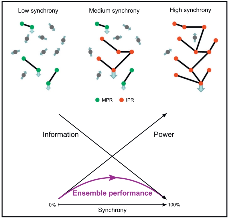
Oscillations form a ubiquitous feature of the central nervous system. Evidence is accruing from cortical and sub-cortical recordings that these rhythms may be functionally important, although the precise details of their roles remain unclear. The basal ganglia share this predilection for rhythmic activity which, as we see in Parkinson's disease, becomes further enhanced in the dopamine depleted state. While certain cortical rhythms appear to penetrate the basal ganglia, others are transformed or blocked. Here, we discuss the functional association of oscillations in the basal ganglia and their relationship with cortical activity. We further explore the neural underpinnings of such oscillatory activity, including the important balance to be struck between facilitating information transmission and limiting information coding capacity. Finally, we introduce the notion that synchronised oscillatory activity can be broadly categorised as immutability promoting rhythms that reinforce incumbent processes, and mutability promoting rhythms that favour novel processing.

Keywords: Basal ganglia; Cross-frequency; Deep brain stimulation; Immutable; Information theory; Parkinson's disease.


Figures

Fig. 1
Fig. 1
Anatomy of the basal ganglia and dopaminergic modulation of basal ganglia oscillations in Parkinson’s disease. [A] Major anatomical connections within and between the basal ganglia and cortex. [B] Schematic of the relationship between dopaminergic activity in the basal ganglia, and beta activity in health and in PD. Upper panel; normal state associated with low levels of beta. Middle panel; untreated PD. Due to the loss of nigral dopaminergic neurones there is less presynaptic dopamine for release in the striatum and STN. Net dopamine, the sum of tonic and phasic release modes, is low and the dynamic range of dopamine variation begins from a lower threshold than in the healthy state. Lower panel; treatment of PD patients with levodopa or dopamine agonists is thought to change the set-point of the system, driving the dynamic range into normal limits. [B] Adapted with permission, Jenkinson and Brown (2011).
Fig. 2
Fig. 2
Grand average spectra from the STN observed ON and OFF dopaminergic medication in a cohort of patients with Parkinson’s disease. [A] Time-evolving spectra centred about the onset of movement during self-initiated wrist-extensions. Beta activity desynchronises prior to and during movement, particularly ON medication. [B] Time-averaged resting power spectrum. Note the reduction in low, but not high frequency beta power ON medication. There are also peaks in the theta/alpha, gamma and high-frequency (250–350 Hz) bands. [A] and [B] adapted with permission, López-Azcárate et al. (2010).
Fig. 3
Fig. 3
Multiplexing and cross-frequency interaction. [A] Functional organisation of a model of the STN in Parkinson’s disease depicting neurons classified by their oscillatory activity. Neurons are described as oscillating at tremor frequency band (TFB), high-frequency [8–20 Hz] band (HFB) or dual-frequency band (DFB). The model involves multiplexing both at the nuclear and neuronal levels. [B] Example of the time–frequency firing pattern of a DFB neuron oscillating simultaneously at both tremor and high (beta-range) frequencies; clear evidence of multiplexing within a neuron. [C] Cross-frequency coupling between theta-phase and gamma-amplitude in the rat striatum obtained during navigation of a T-maze task. [A] and [B] adapted with permission, Moran et al. (2008), [C] adapted with permission, Tort et al. (2008).
Fig. 4
Fig. 4
Neural synchrony and its relationship to information theory. Note drop in information coding capacity (as formally indexed by entropy in C and D) with increased synchronisation (or its surrogate LFP power). Reproduced with permission, Hanslmayr et al. (2012).
Fig. 5
Fig. 5
Idealised relationship between ensemble performance and neural synchrony. As sub-populations of neurons become correlated, the signal-to-noise ratio of that cluster relative to the population increases. At the same time the amount of information that can be transmitted by the ensemble decreases. The result is an inverted U-shape to ensemble performance as synchronisation increases. Mutability promoting rhythms (MPR), such as those in the gamma band, operate to the left of the ensemble performance curve, as dictated by their low power, weakly and locally synchronised nature. Increases in synchronisation in these activities improve ensemble performance and the ability to react to changing circumstances. Immutability promoting rhythms (IPR) such as alpha and beta, tend to operate to the right of the ensemble performance curve in keeping with their higher power, more extensively synchronised nature. This degree of synchronisation is more likely to be supported by recurrent networks where reinforcement of the ongoing oscillations leads to a further reduction in reactivity to external perturbation. Increases in synchronisation in more synchronised immutability promoting rhythms diminish ensemble performance and the ability to react to changing circumstances. This may, in turn, be exaggerated by plastic reconfiguration of networks by the rhythmic activities themselves (see text).

References

    1. Albin RL, Young AB, Penney JB. The functional anatomy of basal ganglia disorders. Trends Neurosci. 1989;12:366–375. - PubMed
    1. Alegre M, Alonso-Frech F, Rodríguez-Oroz MC, Guridi J, Zamarbide I, Valencia M, Manrique M, Obeso JA, Artieda J. Movement-related changes in oscillatory activity in the human subthalamic nucleus: ipsilateral vs. contralateral movements. Eur. J. Neurosci. 2005;22:2315–2324. - PubMed
    1. Alegre M, Lopez-Azcarate J, Obeso I, Wilkinson L, Rodriguez-Oroz MC, Valencia M, Garcia-Garcia D, Guridi J, Artieda J, Jahanshahi M, Obeso JA. The subthalamic nucleus is involved in successful inhibition in the stop-signal task: a local field potential study in Parkinson’s disease. Exp. Neurol. 2013;239:1–12. - PubMed
    1. Alonso-Frech F. Slow oscillatory activity and levodopa-induced dyskinesias in Parkinson’s disease. Brain. 2006;129:1748–1757. - PubMed
    1. Androulidakis AG, Doyle LMF, Yarrow K, Litvak V, Gilbertson TP, Brown P. Anticipatory changes in beta synchrony in the human corticospinal system and associated improvements in task performance. Eur. J. Neurosci. 2007;25:3758–3765. - PubMed

Publication types