. 2013;5(6):1151-64.
doi: 10.1093/gbe/evt084.

Complete genomes of two dipteran-associated spiroplasmas provided insights into the origin, dynamics, and impacts of viral invasion in spiroplasma

Affiliations

Complete genomes of two dipteran-associated spiroplasmas provided insights into the origin, dynamics, and impacts of viral invasion in spiroplasma

Chuan Ku et al. Genome Biol Evol. 2013.

Abstract

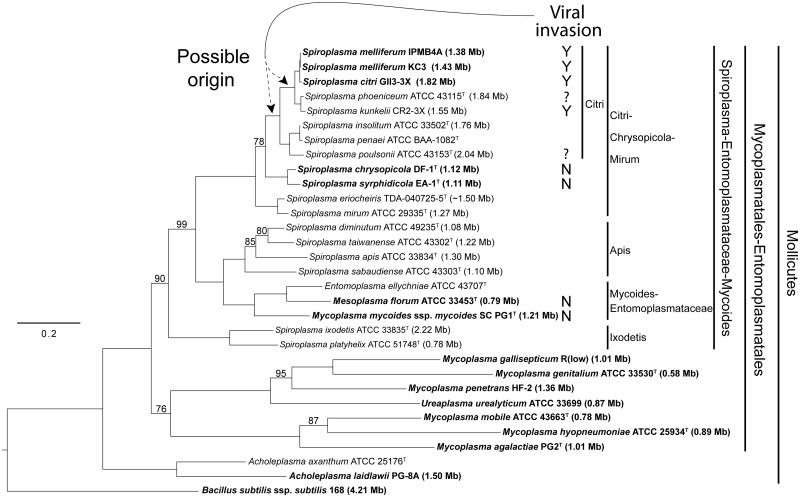
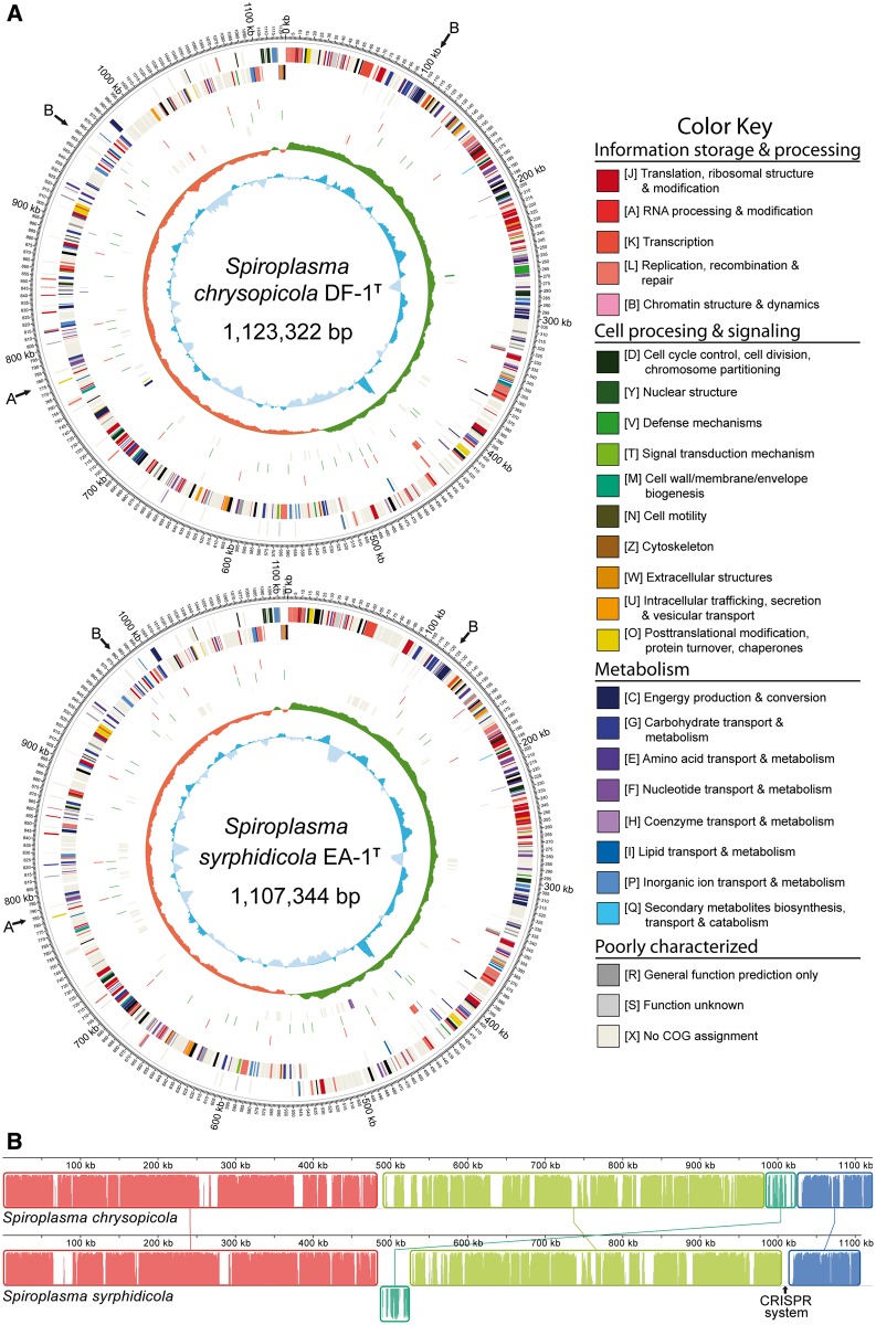
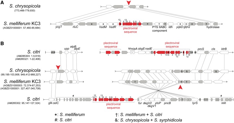
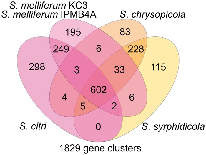
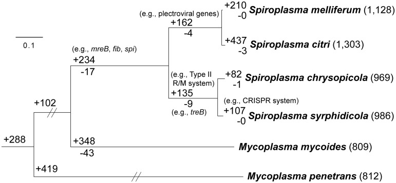
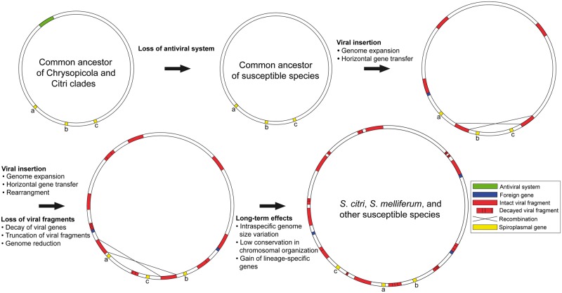
Spiroplasma is a genus of wall-less, low-GC, Gram-positive bacteria with helical morphology. As commensals or pathogens of plants, insects, ticks, or crustaceans, they are closely related with mycoplasmas and form a monophyletic group (Spiroplasma-Entomoplasmataceae-Mycoides) with Mycoplasma mycoides and its relatives. In this study, we report the complete genome sequences of Spiroplasma chrysopicola and S. syrphidicola from the Chrysopicola clade. These species form the sister group to the Citri clade, which includes several well-known pathogenic spiroplasmas. Surprisingly, these two newly available genomes from the Chrysopicola clade contain no plectroviral genes, which were found to be highly repetitive in the previously sequenced genomes from the Citri clade. Based on the genome alignment and patterns of GC-skew, these two Chrysopicola genomes appear to be relatively stable, rather than being highly rearranged as those from the Citri clade. Phylogenetic analyses suggest that the susceptibility to plectroviral invasion probably originated in the common ancestor of the Citri clade or one of its subclades. This susceptibility may be attributed to the absence of antiviral systems found in the Chrysopicola clade. Using the virus-free genomes of the Chrysopicola clade as references, we inferred the putative viral integration sites in the Citri genomes. Comparisons of syntenic regions suggest that the extensive viral invasion in the Citri clade promoted genome rearrangements and expansions. More importantly, the viral invasion may have facilitated horizontal gene transfers that contributed to adaptation in the Citri clade.

Keywords: Citri–Chrysopicola–Mirum clade; Mollicutes; Spiroplasma chrysopicola; Spiroplasma syrphidicola; plectrovirus; viral insertion.


Figures

F<sc>ig</sc>. 1.—
Fig. 1.—
Molecular phylogeny of the Mycoplasmatales–Entomoplasmatales clade. The maximum-likelihood phylogeny was based on 16S rDNA and rpoB sequences, with emphasis on the Citri–Chrysopicola–Mirum clade. All nodes, except where indicated, received 100% bootstrap support. Taxa with genome sequences in GenBank are highlighted in bold, with genome sizes indicated in parentheses. The genome sizes of the species that do not have complete genome sequences available are estimates based on pulsed-field gel electrophoresis. The occurrence of plectroviral fragments in the chromosome is indicated: “N”: no plectroviral fragments in complete genome sequences; “Y”: plectroviral fragments comprising a significant proportion of genome sequences; “?”: presence of plectroviral fragments in the chromosome indicated by Southern hybridization (Spiroplasma phoeniceum) or susceptibility to plectroviral infection (S. poulsonii).
F<sc>ig</sc>. 2.—
Fig. 2.—
Genome maps and alignment of Spiroplasma chrysopicola and S. syrphidicola. (A) Genome maps of S. chrysopicola and S. syrphidicola. Rings from the outermost to the center: 1) scale marks, 2) protein-coding genes on the forward strand, 3) protein-coding genes on the reverse strand, 4) tRNA (purple) and rRNA (red) genes, 5) putative sites of viral insertions in S. melliferum KC3 (red) relative to the respective genomes, 6) putative sites of viral insertions in S. citri (green) relative to the respective genomes, 7) protein-coding genes unique to the respective genomes, 8) GC skew, and 9) GC content. Protein-coding genes are color coded according to their COG categories. The positions of the genomic regions shown in figure 3A and B are indicated by arrows. (B) Whole-genome alignment between S. chrysopicola and S. syrphidicola.
F<sc>ig</sc>. 3.—
Fig. 3.—
Examples of syntenic regions with plectroviral genes in the Citri clade. Annotated plectroviral genes are highlighted in red with labels indicating the ORF number assignments. Hypothetical genes with unknown functions are in gray. Genes within plectroviral sequences, where not indicated, are found in both Spiroplasma citri and S. melliferum (†). Putative viral insertion positions are indicated by red arrowheads. The corresponding regions in S. syrphidicola contain the same genes in the same order as in S. chrysopicola and are not shown for simplicity. In (A), the corresponding region in S. citri has other plectroviral fragments in the vicinity (fig. 2) and is scattered into different contigs.
F<sc>ig</sc>. 4.—
Fig. 4.—
Numbers of shared and unique gene clusters among Spiroplasma chrysopicola, S. syrphidicola, S. citri, and S. melliferum. Genomes of the two S. melliferum strains (IPMB4A and KC3) were analyzed as a single pan-genome of S. melliferum. For detailed lists of these orthologous gene clusters, see supplementary table S2, Supplementary Material online.
F<sc>ig</sc>. 5.—
Fig. 5.—
Phylogenetic distribution pattern of homologous gene clusters. The organismal phylogeny is inferred from the concatenated protein alignment of 267 single-copy genes shared by all species (with 106,639 aligned amino acid sites). All internal nodes received 100% bootstrap support based on 1,000 resampling and maximum likelihood inference. The numbers in parentheses after species names indicate the number of gene clusters found in each species. The numbers above a branch and preceded by a “+” sign indicate the number of homologous gene clusters that are uniquely present in all daughter lineages; the numbers below a branch and preceded by a “−” sign indicate the number of homologous gene clusters that are uniquely absent. For example, 135 gene clusters are shared by Spiroplasma chrysopicola and S. syrphidicola and do not contain any homolog from any of the other four species compared; similarly, nine gene clusters are missing in these two Spiroplasma species but are present in all other four species. For detailed lists of these orthologous gene clusters, see supplementary table S3, Supplementary Material online.
F<sc>ig</sc>. 6.—
Fig. 6.—
A model for the genome evolution in spiroplasmas susceptible to viral invasion.

References

    1. Alexeev D, et al. Application of Spiroplasma melliferum proteogenomic profiling for the discovery of virulence factors and pathogenicity mechanisms in host-associated spiroplasmas. J Proteome Res. 2012;11:224–236. - PubMed
    1. Altschul SF, et al. Gapped BLAST and PSI-BLAST: a new generation of protein database search programs. Nucleic Acids Res. 1997;25:3389–3402. - PMC - PubMed
    1. André A, Maccheroni W, Doignon F, Garnier M, Renaudin J. Glucose and trehalose PTS permeases of Spiroplasma citri probably share a single IIA domain, enabling the spiroplasma to adapt quickly to carbohydrate changes in its environment. Microbiology. 2003;149:2687–2696. - PubMed
    1. Bai X, et al. Living with genome instability: the adaptation of phytoplasmas to diverse environments of their insect and plant hosts. J Bacteriol. 2006;188:3682–3696. - PMC - PubMed
    1. Bai X, Hogenhout SA. A genome sequence survey of the mollicute corn stunt spiroplasma Spiroplasma kunkelii. FEMS Microbiol Lett. 2002;210:7–17. - PubMed

Publication types

Associated data