. 2013 May 28:9:667.
doi: 10.1038/msb.2013.24.

The Neurospora photoreceptor VIVID exerts negative and positive control on light sensing to achieve adaptation

Affiliations

The Neurospora photoreceptor VIVID exerts negative and positive control on light sensing to achieve adaptation

Elan Gin et al. Mol Syst Biol. .

Abstract

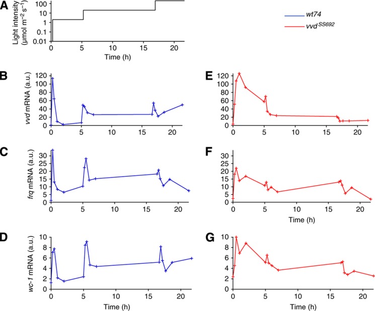
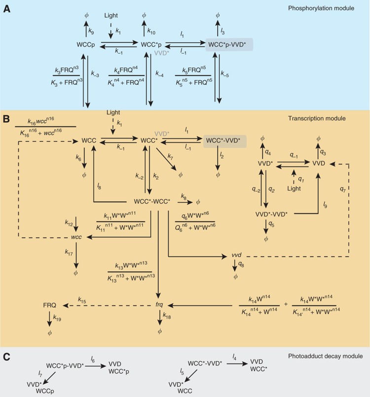
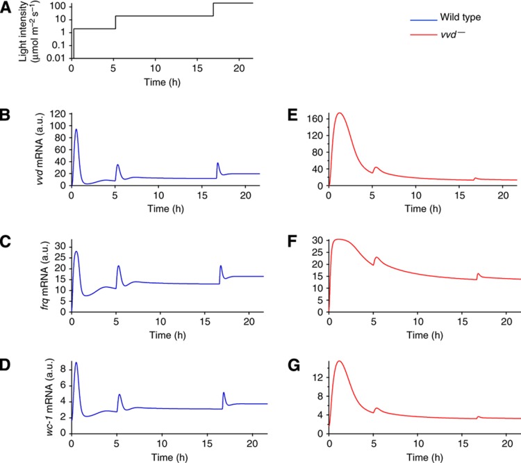
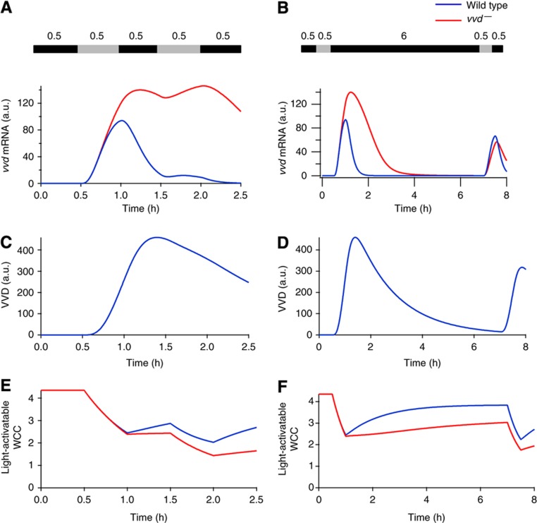
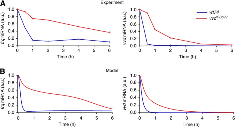
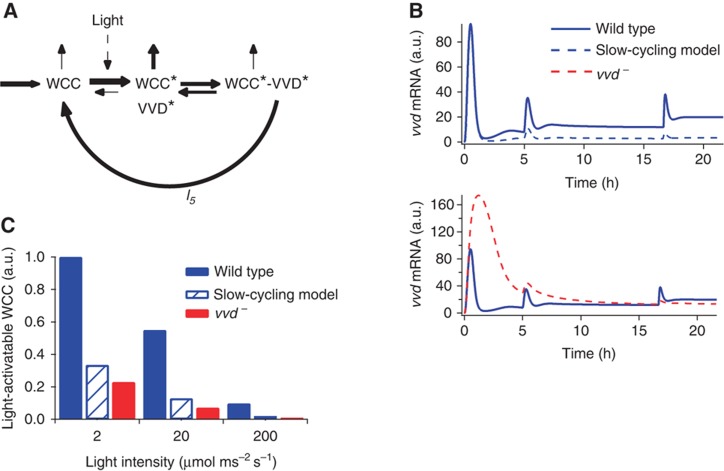
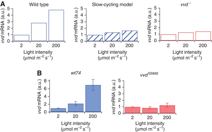
The light response in Neurospora is mediated by the photoreceptor and circadian transcription factor White Collar Complex (WCC). The expression rate of the WCC target genes adapts in daylight and remains refractory to moonlight, despite the extraordinary light sensitivity of the WCC. To explain this photoadaptation, feedback inhibition by the WCC interaction partner VIVID (VVD) has been invoked. Here we show through data-driven mathematical modeling that VVD allows Neurospora to detect relative changes in light intensity. To achieve this behavior, VVD acts as an inhibitor of WCC-driven gene expression and, at the same time, as a positive regulator that maintains the responsiveness of the photosystem. Our data indicate that this paradoxical function is realized by a futile cycle that involves the light-induced sequestration of active WCC by VVD and the replenishment of the activatable WCC pool through the decay of the photoactivated state. Our quantitative study uncovers a novel network motif for achieving sensory adaptation and defines a core input module of the circadian clock in Neurospora.


Conflict of interest statement

The authors declare that they have no conflict of interest.

Figures

Figure 1
Figure 1
Rapid adaptation and repeated induction depend on VVD. Light induction experiments for vvd, wc-1 and frq. Liquid cultures of the vvd loss-of-function mutant (vvdSS69) and its corresponding wild-type strain (wt74) were raised in light for 24 h and then transferred to constant darkness for 24 h before light induction. Initial light intensity was 2 μmol m−2 s−1 and was raised to 20 μmol m−2 s−1 after 300 min and again to 200 μmol m−2 s−1 after 1000 min (A). Samples were collected at the indicated time points and mRNA levels for vvd, frq and wc-1 were measured via qRT–PCR. The wild-type vvd mRNA levels at t=0 were set to 1. To compare the relative levels of the mRNAs, we corrected for the efficiency of the various RT–PCR probes used, by performing qPCR with a dilution series of genomic DNA. The values were corrected with reference to vvd (further details in the Supplementary Information). Wild-type data are shown in panels (BD) and vvdSS692 mutant data are shown in panels (EG). Throughout the paper, wild-type data are shown in blue and vvdSS692 mutant data are shown in red. Source data for this figure is available on the online supplementary information page.
Figure 2
Figure 2
Model scheme. Protein variables are indicated in normal font and mRNA variables are italicized. Light-activated forms of protein are denoted with an asterix. Phosphorylated states are of the form WCCp. The model is described in three modules: a phosphorylation module (A, shaded blue) involving FRQ-dependent phosphorylation of the WCC; a transcription module (B) that includes the light activation of WCC, subsequent homodimerization and transcription; and the photoadduct decay module (C, shaded gray). The photoadduct decay module shows the additional pathways for breakdown of the heterodimer complex.
Figure 3
Figure 3
The model accounts for the photoresponse dynamics of the wild type and mutant. The model scheme was fitted to the light induction experiments with the light regime shown in (A), and, consistent with the experimental data, the wild type shows repeated activation in response to increasing light (BD), while responsiveness is diminished in the mutant (EG).
Figure 4
Figure 4
VVD sets the refractory period after light response. Model-simulated responses to different dark/light regimes with light levels of 20 μmol m−2 s−1. The intervening dark incubation period between the two light pulses are 30 min (left panel) or 6 h (right panel). (A and B) After only 30 min of intervening darkness, the vvd mutant responds to the second light pulse, whereas the wild type requires a period of 6 h in the dark before it can respond again. (C and D) A substantial pool of VVD still remains after 30 min in the dark, thus preventing a response to the second light pulse in the wild type. After 6 h, the VVD pool is depleted, restoring full responsiveness. (E and F) Light-activatable WCC (dark form) levels recover faster in the wild type than the vvd mutant during both durations of dark period, illustrating the role of VVD in restoring the WCC. On exposure to the second light pulse, the WCC is activated in both the wild type and vvd mutant. However, the VVD remaining after 30 min of dark suppresses the light response, and only after 6 h of darkness are VVD levels low such that the wild type is able to respond to the light pulse.
Figure 5
Figure 5
VVD represses transcriptional activity after transfer to dark. (A) Repression of frq and vvd RNA synthesis upon light–dark transfer is slow in vvdSS692. Cultures of wt and vvdSS692 were grown in constant light (20 μmol m−2 s−1) for 2 days, transferred to darkness and collected at the indicated time points. Levels of vvd and frq mRNA are shown as determined by real-time PCR. mRNA levels are normalized to the value at the light-to-dark transfer point (t=0). (B) Simulations reproduce the experimental result showing frq and vvd mRNA levels elevated for up to 4 h in the vvd mutant and wild-type levels reaching baseline within 1 h. Source data for this figure is available on the online supplementary information page.
Figure 6
Figure 6
Photoadduct decay constitutes a futile cycle that ensures repeated sensitivity. (A) Schematic representation of the futile cycling arising from photoadduct decay of the WCC–VVD hetereodimer. Thickness of the arrows indicate the relative rate of reactions. (B) Model simulations show responsiveness to increasing light stimuli is maintained via the futile cycle. Upper panel: the rate of photoadduct decay, l5, is decreased by a factor of 10. The first response is identical to the wild type (solid blue) but the system loses responsiveness to increasing light stimuli (dashed blue), while still functioning as an inhibitor of light-mediated WCC activity. Lower panel: the vvd mutant loses both the ability to respond to increasing light (similar to the slow-cycling model) and downregulation of the response in constant stimulus. (C) Repeated responsiveness is maintained by a sizeable pool of light-activatable WCC (WCC). Simulations show wild-type levels are higher than the vvd mutant and the slow-cycling model.
Figure 7
Figure 7
VVD tracks ambient light levels. (A) Model simulations show wild-type vvd mRNA levels rise up to fivefold with increasing light intensity. The simulated levels from both the slow-cycling model and the vvd mutant remain constant over the three light intensities. All levels are normalized to the respective mRNA values at 2 μmol m−2 s−1. (B) Experimental results confirm the model simulations. Neurospora cultures of the indicated strains were grown for 48 h in constant light at 2, 20 and 200 μmol m−2 s−1, respectively. Samples were collected and mRNA was measured via qRT–PCR. Results shown are from at least two independent experiments measured in triplicates. Source data for this figure is available on the online supplementary information page.

References

    1. Akman OE, Locke JC, Tang S, Carré I, Millar AJ, Rand DA (2008) Isoform switching facilitates period control in the Neurospora crassa circadian clock. Mol Syst Biol 4: 164. - PMC - PubMed
    1. Akman OE, Rand DA, Brown PE, Millar AJ (2010) Robustness from flexibility in the fungal circadian clock. BMC Syst Biol 4: 1–17 - PMC - PubMed
    1. Arpaia G, Cerri F, Baima S, Macino G (1999) Involvement of protein kinase C in the response of Neurospora crassa to blue light. Mol Gen Genet 262: 314–322 - PubMed
    1. Baker CL, Loros JJ, Dunlap JC (2012) The circadian clock of Neurospora crassa. FEMS Microbiol Rev 36: 95–110 - PMC - PubMed
    1. Behar M, Hao N, Dohlman HG, Elston TC (2007) Mathematical and computational analysis of adaptation via feedback inhibition in signal transduction pathways. Biophys J 93: 806–821 - PMC - PubMed

Publication types

MeSH terms

LinkOut - more resources