. 2013 Jun 3;210(6):1189-200.
doi: 10.1084/jem.20130392. Epub 2013 May 27.

Differentiation of CD8 memory T cells depends on Foxo1

Affiliations

Differentiation of CD8 memory T cells depends on Foxo1

Rodrigo Hess Michelini et al. J Exp Med. .

Abstract

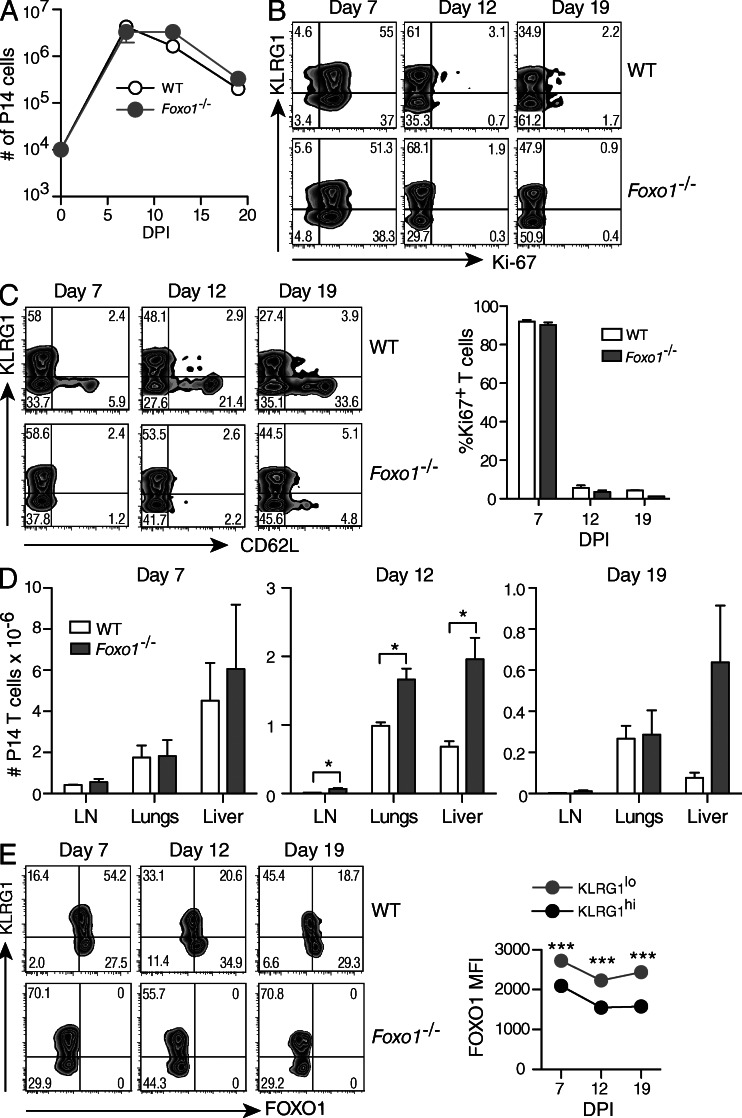
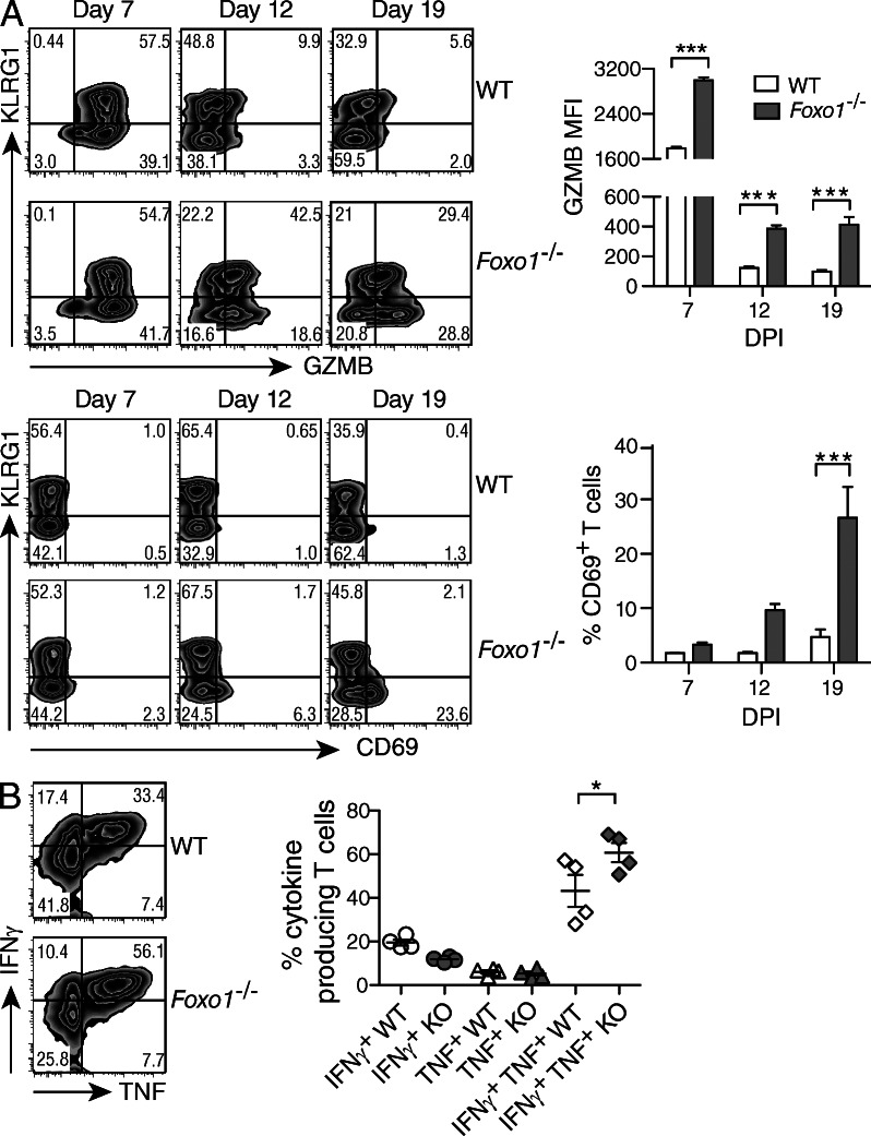
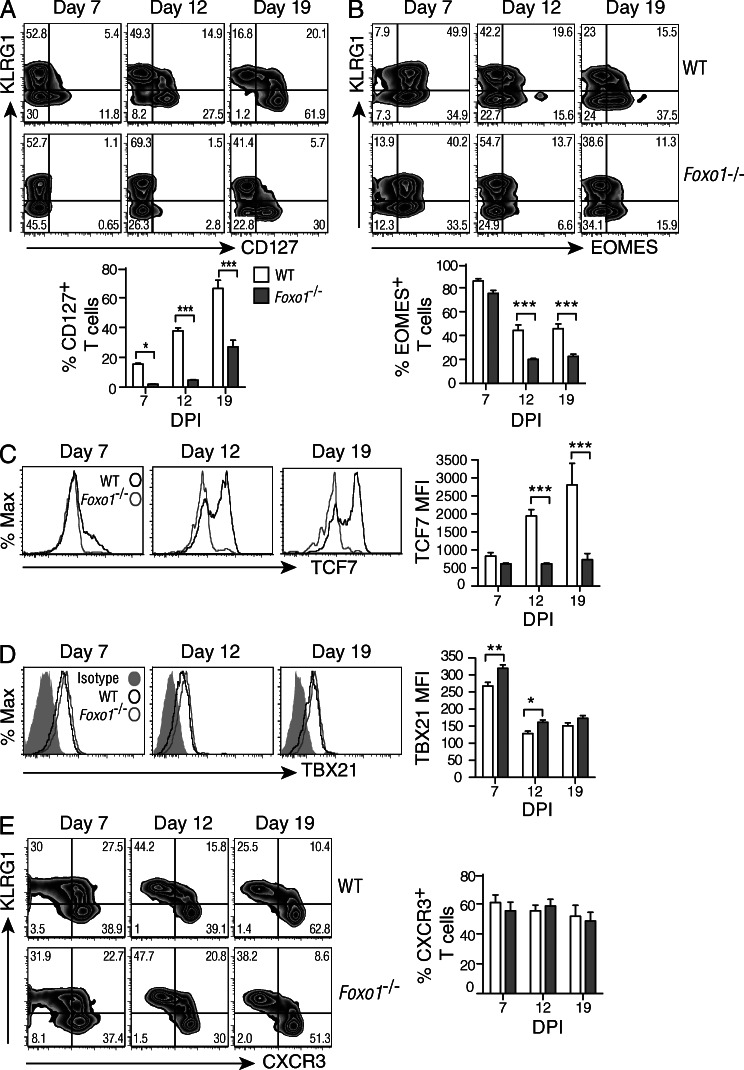
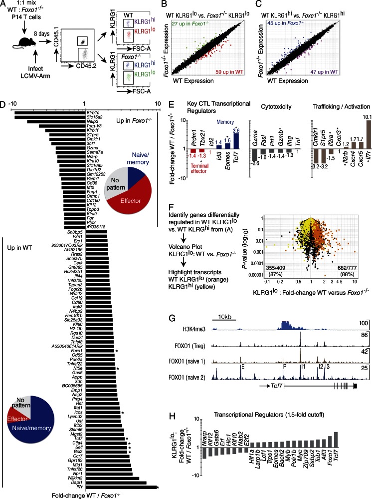
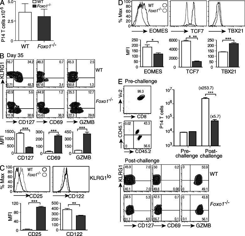
The forkhead O transcription factors (FOXO) integrate a range of extracellular signals, including growth factor signaling, inflammation, oxidative stress, and nutrient availability, to substantially alter the program of gene expression and modulate cell survival, cell cycle progression, and many yet to be unraveled cell type-specific responses. Naive antigen-specific CD8(+) T cells undergo a rapid expansion and arming of effector function within days of pathogen exposure. In addition, by the peak of expansion, they form precursors to memory T cells capable of self-renewal and indefinite survival. Using lymphocytic choriomeningitis virus Armstrong to probe the response to infection, we found that Foxo1(-/-) CD8(+) T cells expand normally with no defects in effector differentiation, but continue to exhibit characteristics of effector T cells long after antigen clearance. The KLRG1(lo) CD8(+) T cells that are normally enriched for memory-precursor cells retain Granzyme B and CD69 expression, and fail to up-regulate TCF7, EOMES, and other memory signature genes. As a correlate, Foxo1(-/-) CD8(+) T cells were virtually unable to expand upon secondary infection. Collectively, these results demonstrate an intrinsic role for FOXO1 in establishing the post-effector memory program that is essential to forming long-lived memory cells capable of immune reactivation.


Figures

Figure 1.
Figure 1.
Kinetics of CD8+ T cell expansion in response to LCMV-Arm infection. A 1:1 mix of 104 CD45.1+/2+ WT (Foxo1f/fGZMBCre) and CD45.2+ Foxo1−/− (Foxo1f/fGZMBCre+) P14 cells were transferred to CD45.1+ WT host mice. Mice were then infected with LCMV-Arm, and lymphoid and nonlymphoid organs were harvested and analyzed at day 7, 12, and 19 after infection. (A) WT and Foxo1−/− P14 cells were identified in the spleen by their expression of CD45.1, CD45.2, CD8, and Vα2 and total numbers of P14 cells were determined. DPI, days post infection. (B) Ki67 staining of spleen cells from WT and Foxo1−/− P14 cells at different time points. (C) Expression of KLRG1 and CD62L on WT and Foxo1−/− P14 cells isolated from the spleen. (D) Total numbers of WT and Foxo1−/− P14 cells in the indicated organs. (E) Expression of KLRG1 and FOXO1 on WT and Foxo1−/− P14 cells isolated from the spleen. Data representative of 1 out of 3 independent experiments each, with n = 3,4.
Figure 2.
Figure 2.
Phenotype of virus-specific CD8+ T cells. A 1:1 mix of 104 CD45.1+/2+ WT (Foxo1f/fGZMBCre) and CD45.2+ Foxo1−/− (Foxo1f/fGZMBCre+) P14 cells were transferred to CD45.1+ WT host mice. Mice were then infected with LCMV-Arm, and spleen cells were harvested and analyzed at different time points. (A) The expression of KLRG1, GZMB, and CD69 was determined in WT and Foxo1−/− P14 cells at different time points (left), and the amount of GZMB (MFI) and percentage of CD69 are depicted (right). Data are representative of 3 independent experiments, with n = 3–4. (B) At day 7 after infection, spleen cells were stimulated with PMA and ionomycin and the frequency of IFN-γ– and TNF-producing cells was determined. Data are representative of 2 independent experiments each, with n = 4.
Figure 3.
Figure 3.
Enhanced effector phenotype in the absence of FOXO1. A 1:1 mix of 1 × 10⁴ CD45.1+/2+ WT (Foxo1f/fGZMBCre) and CD45.2+ Foxo1−/− (Foxo1f/fGZMBCre+) P14 cells were transferred to CD45.1+ WT host mice. Mice were then infected with LCMV-Arm, and spleen cells were harvested and analyzed at different time points. (A–E) WT, open bars; Foxo1−/−, filled bars. (A) The expression of KLRG1 and CD127 was determined in WT and Foxo1−/− P14 cells, and the percentage of CD127+ cells is depicted (bottom). (B) The expression of KLRG1 and EOMES was determined in WT and Foxo1−/− P14 cells, and the percentage of EOMES+ cells is depicted (bottom). (C and D) WT and Foxo1−/− P14 cells were analyzed for their expression of TCF7 (C) and TBX21 (D), and the amount of both are presented as mean fluorescence intensity (MFI, right). (E) The expression of KLRG1 and CXCR3 was determined in WT and Foxo1−/− P14 cells (left), and the percentage of CXCR3+ cells depicted (right). Data are representative of 3 independent experiments each, with n = 3–4.
Figure 4.
Figure 4.
Deletion of Foxo1 prevents the development of a memory program of gene expression. A 1:1 mix of 104 WT (Foxo1f/f Rosa26) and Foxo1−/− (Foxo1f/f Rosa26Cre-ERT2) P14 cells were prepared from tamoxifen-treated mice and transferred to CD45.1+ WT host mice. Microarray gene-expression analysis was performed by sorting WT and Foxo1−/− KLRG1lo P14 cells 8 d after infection with LCMV-Arm. (A) Experimental scheme for mixed transfer and FACS of WT and Foxo1−/− P14 T cells on day 8 of LCMV-Armstrong infection. The four double-sorted populations were analyzed on the same microarray chip. (B) KLRG1lo WT versus Foxo1−/− reveals 59 genes up and 27 genes down with a twofold cutoff. (C) KLRG1hi WT versus Foxo1−/− reveals 47 genes up and 45 genes down with a twofold cutoff. (D) Identification of genes KLRG1lo WT / Foxo1−/− with a twofold cutoff. Pie charts indicate portion of genes with an expression pattern tied to naive/memory (blue), effector (red), or no pattern (gray). Asterisk indicates expression was verified by flow cytometry. (E) Classification of a subset of the genes in (D) by function. (F) Genes differentially regulated in WT KLRG1lo versus WT KLRG1hi, were overlaid with genes from KLRG1lo WT versus KLRG1lo Foxo1−/− CD8+ T cells, showing nearly a 90% concordance of transcripts. (G) Naive CD4+ ChIP-Seq data showing several FOXO1 binding sites proximal to and within Tcf7. Data were analyzed from T reg cells and two different datasets analyzing naive CD4 T cells. Peaks are labeled as putative: intergenic enhancer (E), promoter (P), and intronic enhancers 1, 2, and 3 (I1, I2, I3). Peak sequences are listed in Table I. (H) Transcription factors known to play a role in the formation of memory cells, summarizing data presented in D with the addition of other transcription factors differentially regulated in WT vs. Foxo1−/− P14 cells. Data are from 1 experiment with n = 3.
Figure 5.
Figure 5.
Foxo1−/− CD8+ T cells fail to expand upon secondary viral challenge. (A–E) WT, open squares; Foxo1−/− filled squares. A 1:1 mix of 104 CD45.1+/2+ WT (Foxo1f/fGZMBCre) and CD45.2/2+ Foxo1−/− (Foxo1f/fGZMBCre+) P14 cells were prepared and transferred to CD45.1+ WT host mice. Mice were then infected with LCMV-Arm, and spleen cells were analyzed 35 d after infection. (A) Number of cells recovered per spleen at day 35. (B) WT and Foxo1−/− P14 cells were analyzed by their expression of KLRG1 and CD127, CD69, and GZMB (top), and the MFI of these molecules was quantified on the KLRG1lo population (bottom). (C) Expression of CD25 and CD122 on KLRG1lo P14 cells. (D) WT and Foxo1−/− P14 cells were analyzed for expression of transcription factors important for CD8+ memory and effector T cells, and the amount of protein expressed on the KLRG1lo population graphed as MFI. (E) P14 (CD8+Vα2+) WT (CD45.1+/2+) and Foxo1−/− (CD45.1+) cells from hosts 35 d after LCMV-Arm infection were sorted and transferred at a 1:1 ratio into naive WT mice. Mice were then infected with LCMV-Arm and the expansion of WT and Foxo1−/− P14 cells determined at d 5 after infection (top right). The phenotype of P14 T cells 5 d after secondary challenge (bottom). Data are representative of 2 independent experiments for a total n = 11.

References

    1. Almeida M. 2011. Unraveling the role of FoxOs in bone—insights from mouse models. Bone. 49:319–327 10.1016/j.bone.2011.05.023 - DOI - PMC - PubMed
    1. Almeida M., Han L., Martin-Millan M., O’Brien C.A., Manolagas S.C. 2007. Oxidative stress antagonizes Wnt signaling in osteoblast precursors by diverting beta-catenin from T cell factor- to forkhead box O-mediated transcription. J. Biol. Chem. 282:27298–27305 10.1074/jbc.M702811200 - DOI - PubMed
    1. Araki K., Turner A.P., Shaffer V.O., Gangappa S., Keller S.A., Bachmann M.F., Larsen C.P., Ahmed R. 2009. mTOR regulates memory CD8 T-cell differentiation. Nature. 460:108–112 10.1038/nature08155 - DOI - PMC - PubMed
    1. Bachmann M.F., Wolint P., Walton S., Schwarz K., Oxenius A. 2007. Differential role of IL-2R signaling for CD8+ T cell responses in acute and chronic viral infections. Eur. J. Immunol. 37:1502–1512 10.1002/eji.200637023 - DOI - PubMed
    1. Banerjee A., Gordon S.M., Intlekofer A.M., Paley M.A., Mooney E.C., Lindsten T., Wherry E.J., Reiner S.L. 2010. Cutting edge: The transcription factor eomesodermin enables CD8+ T cells to compete for the memory cell niche. J. Immunol. 185:4988–4992 10.4049/jimmunol.1002042 - DOI - PMC - PubMed

Publication types

MeSH terms