. 2013 Jun 3;210(6):1265-81.
doi: 10.1084/jem.20122753. Epub 2013 May 27.

A hemolytic pigment of Group B Streptococcus allows bacterial penetration of human placenta

Affiliations

A hemolytic pigment of Group B Streptococcus allows bacterial penetration of human placenta

Christopher Whidbey et al. J Exp Med. .

Abstract

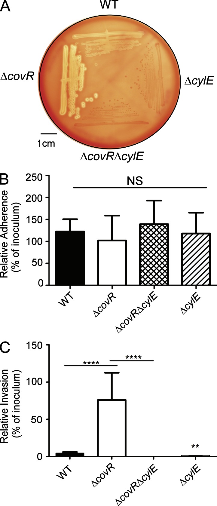
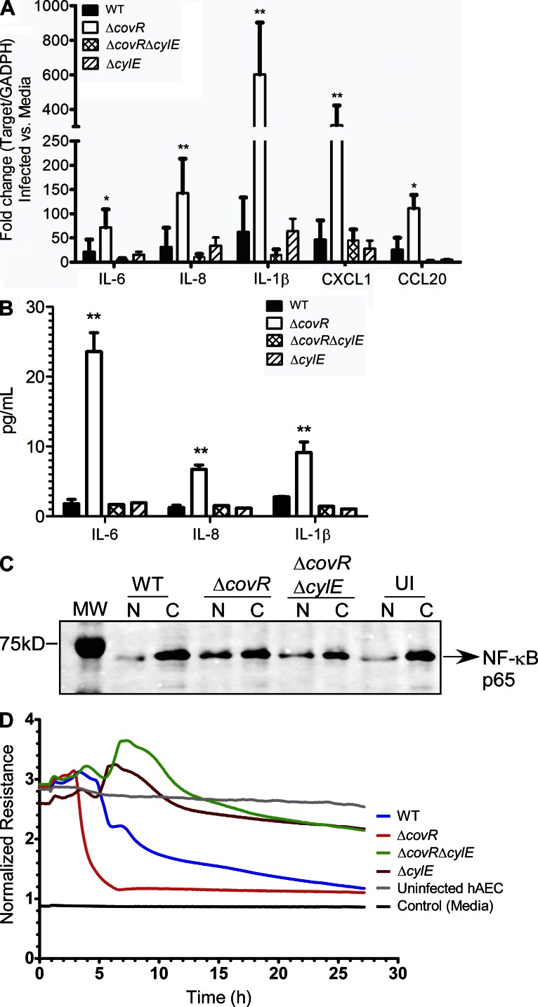
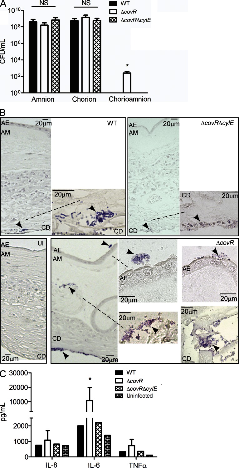
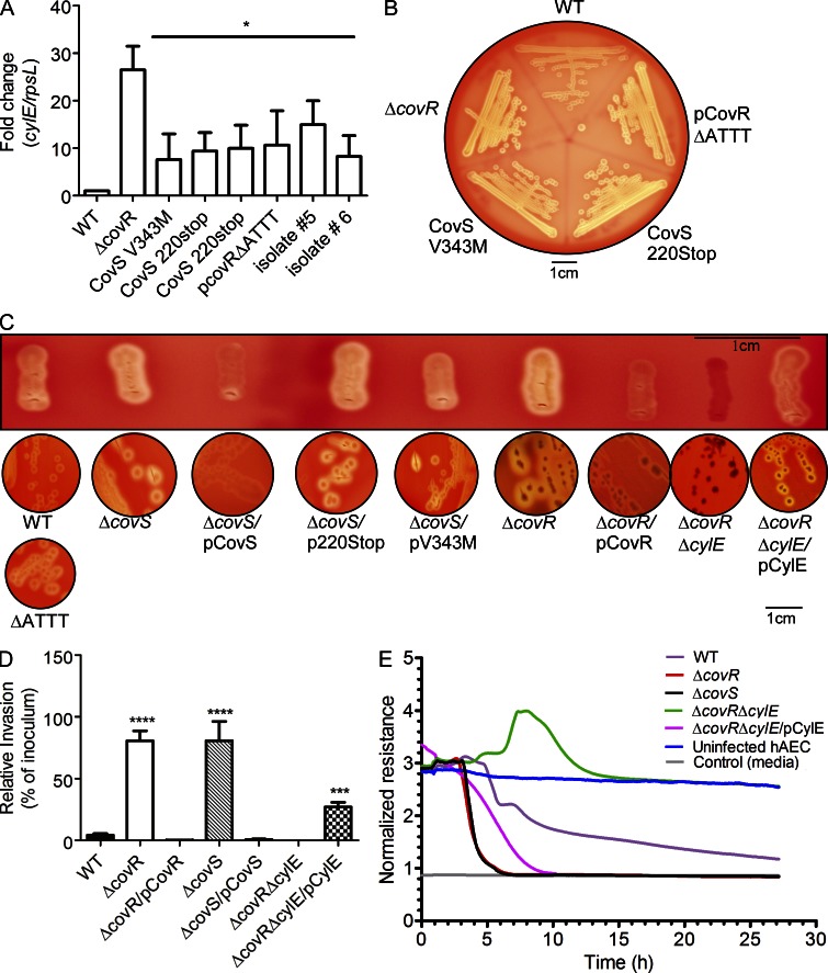
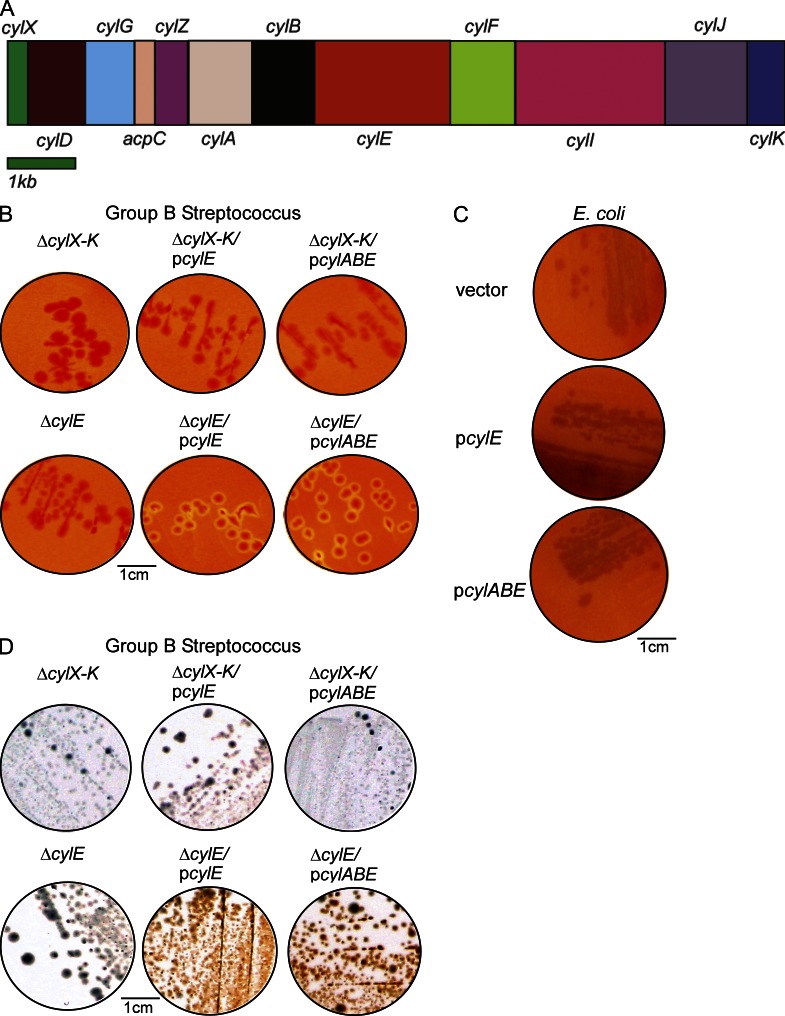
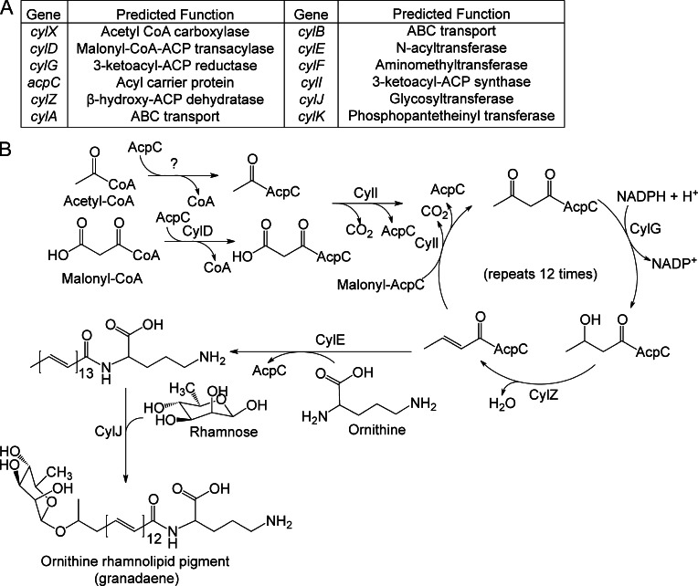
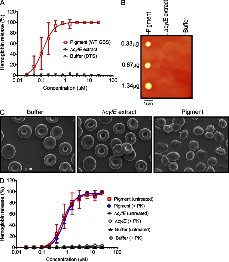
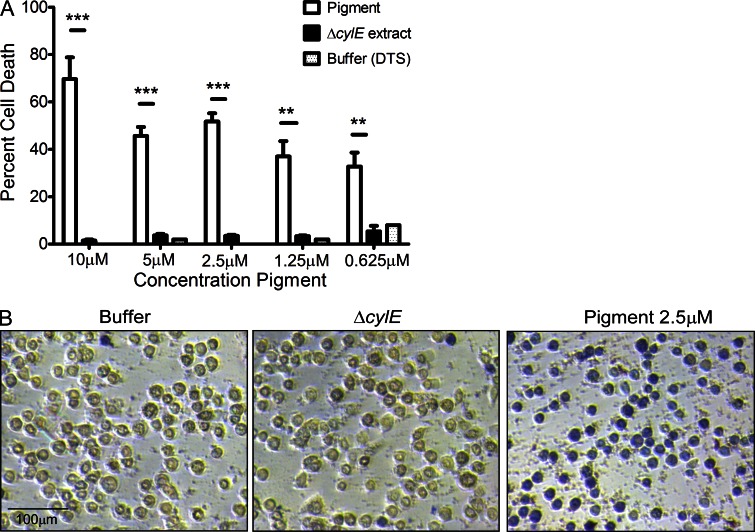
Microbial infection of the amniotic fluid is a significant cause of fetal injury, preterm birth, and newborn infections. Group B Streptococcus (GBS) is an important human bacterial pathogen associated with preterm birth, fetal injury, and neonatal mortality. Although GBS has been isolated from amniotic fluid of women in preterm labor, mechanisms of in utero infection remain unknown. Previous studies indicated that GBS are unable to invade human amniotic epithelial cells (hAECs), which represent the last barrier to the amniotic cavity and fetus. We show that GBS invades hAECs and strains lacking the hemolysin repressor CovR/S accelerate amniotic barrier failure and penetrate chorioamniotic membranes in a hemolysin-dependent manner. Clinical GBS isolates obtained from women in preterm labor are hyperhemolytic and some are associated with covR/S mutations. We demonstrate for the first time that hemolytic and cytolytic activity of GBS is due to the ornithine rhamnolipid pigment and not due to a pore-forming protein toxin. Our studies emphasize the importance of the hemolytic GBS pigment in ascending infection and fetal injury.


Figures

Figure 1.
Figure 1.
Hemolysin promotes GBS invasion of hAECs. (A) Hemolytic activity shown by the zone of clearing around the colonies on sheep blood agar of GBS WT, ΔcovR, and isogenic mutants. A representative image from one of three independent experimental replicates is shown. (B and C) Primary hAECs were isolated from chorioamniotic membranes and adherence and invasion of GBS WT, isogenic ΔcovR, ΔcovRΔcylE, and ΔcylE mutants were compared. Percent adherence (B) and invasion (C) is normalized to that of the initial inoculum. Data shown are the mean and SD obtained from hAECs that were isolated from four independent placentas, and each experiment was performed in triplicate (NS, P > 0.2; ****, P < 0.0001; **, P = 0.008, Student’s t test, error bars ± SD).
Figure 2.
Figure 2.
Hemolysin increases expression of inflammatory mediators and induces barrier disruption in amniotic epithelial cells. (A) qRT-PCR was performed of indicated cytokines/chemokines on RNA isolated from hAEC infected with either WT GBS COH1 or isogenic ΔcovR, ΔcovRΔcylE, and ΔcylE mutants at 4 h after infection. Data shown are the mean and SD obtained from hAECs that were isolated from three independent placentas, performed in triplicate (n = 3; **, P = 0.007; *, P = 0.03, Student’s t test, error bars ± SD). (B) Luminex bead assays were performed on supernatants of hAEC infected with WT GBS or isogenic ΔcovR, ΔcovRΔcylE, and ΔcylE mutants at 4 h after infection. The experiment was performed using hAECs that were isolated from three independent placentas, performed in triplicate (n = 3; **, P < 0.005, Student’s t test, error bars ± SD). (C) Western blots were performed on nuclear (N) and cytoplasmic (C) proteins from GBS-infected hAEC using antibody to NF-κB. Uninfected (UI) hAECs were included as controls. MW = molecular weight marker. A representative image from one of three independent experimental replicates is shown. (D) Barrier resistance of hAEC was monitored in real time using ECIS. A representative image from one of three independent experimental replicates is shown.
Figure 3.
Figure 3.
Hyper-hemolytic GBS penetrate human chorioamnion. Intact chorioamniotic membranes as well as either chorion alone or amnion alone (n = 6) were mounted on a transwell system and infected with either WT GBS COH1 or isogenic ΔcovR and ΔcovRΔcylE strains for a period of 24 h. Aliquots of the media from the lower chamber were analyzed for CFU and cytokines expression (see A–C). (A) GBS penetration of the chorion, amnion, or chorioamniotic membranes (n = 6; NS, P > 0.3; *, P = 0.02, Mann Whitney test, error bars ± SD). (B) Infected chorioamniotic membranes (n = 6) were fixed, embedded, sectioned, and stained using Gram-Tworts stain. A representative placenta where bacteria were not recovered from the lower chamber for any GBS strain is shown. AE, amniotic epithelium; AM, amniotic mesoderm; CD, choriodecidua; UI, uninfected chorioamnion. Arrows represent gram-positive bacteria, and dashed line shows region under higher magnification. (C) ELISA assays on media obtained from the lower chamber of chorioamniotic membranes infected with GBS WT, ΔcovR, and ΔcovRΔcylE (n = 6; *, P = 0.02, Mann Whitney test, error bars ± SD).
Figure 4.
Figure 4.
GBS clinical isolates from women in preterm labor exhibit increased hemolysis and some are associated with CovR/S mutations. (A) qRT-PCR on RNA isolated from log phase GBS (O.D600nm = 0.3). Data are normalized to relative expression of the house keeping gene rpsL and are the mean and SD from five independent biological replicates performed in triplicate (n = 5; *, P = 0.03, Wilcoxon matched-pairs rank test). (B) Hemolytic activity of GBS clinical isolates associated with preterm labor. A representative image from one of three independent experimental replicates is shown. (C) Single colonies were patched and streaked on blood agar. Hemolysis of GBS WT, ΔcovS, ΔcovR, and complementing clones including plasmids encoding CovS220stop and CovSV343M. Hemolysis of GBS with the pCovR promoter deletion compared with WT. Complementation of GBSΔcovRΔcylE with pCylE on hemolytic activity. A representative image from one of three independent experimental replicates is shown. (D) Invasiveness of GBSΔcovS to hAEC compared with WT and ΔcovR. Complementation of ΔcovS and ΔcovR on amniotic invasion and effect of pCylE on amniotic epithelial invasion of ΔcovRΔcylE. Data shown are the mean and SD obtained using hAEC from three independent placentas, performed in triplicate (n = 3; ****, P < 0.0001; ***, P = 0.0006, Student’s t test, error bars ± SD). (E) Barrier disruption of amniotic epithelium by GBS WT, ΔcovS, ΔcovR, and ΔcovRΔcylE. Complementation of GBSΔcovRΔcylE with pCylE on amniotic barrier disruption. A representative image from one of three independent experimental replicates is shown.
Figure 5.
Figure 5.
CylE is necessary but not sufficient for GBS hemolysis. (A) The GBS cyl operon encoding genes cylX-K is shown. (B and C) Complementation of the nonhemolytic GBSΔcylE and ΔcylX-K with plasmids encoding CylE or CylE along with the ABC transporter CylA/B on hemolytic activity. A representative image from one of three independent experimental replicates is shown. (D) Hemolytic and nonhemolytic GBS strains on Granada Media. A representative image from one of three independent experimental replicates is shown.
Figure 6.
Figure 6.
Proposed biosynthetic pathway for the GBS pigment granadaene. (A and B) Predicted functions of the cyl operon proteins and proposed biosynthetic pathway for synthesis of the GBS pigment using acetyl-CoA, malonyl-CoA, ornithine, and rhamnose. CylD conjugates the elongating malonyl CoA units to the acyl carrier protein (ACP) AcpC. CylI links the malonyl CoA to an initial fatty acid–ACP complex, beginning the fatty acid biosynthesis-like pathway. CylG reduces the 3-keto group to a hydroxyl group, which is further reduced to an alkene by CylZ. The cyl operon lacks an enoyl-ACP reductase thereby eliminating the final reduction of the alkene to an alkane. The unsaturated fatty acid serves as a substrate for further elongation by CylI, accounting for the large degree of unsaturation in the pigment. After 13 total rounds of elongation, the fatty acid is conjugated to ornithine by CylE, and glycosylated by CylJ. CylX, CylF, and CylK likely function upstream or downstream of this pathway. CylX is homologous to a component of the acetyl-CoA carboxylase, which generates malonyl-CoA. CylF is an aminomethyltransferase, likely involved in production of the methylated derivative seen in the mass spectrum (Fig. S2). CylK is a putative phosphopantetheinyl transferase, which is involved in the activation of acyl carrier proteins. Although GBS has a separate fatty acid biosynthesis (fab) operon, deletion of genes in the cyl operon (ΔcylX-K, Fig. 5B) abolishes pigment biosynthesis suggesting that the fab and cyl operons are not functionally redundant.
Figure 7.
Figure 7.
The functional basis of GBS hemolytic activity is the pigment. (A) Pigment was added to hRBCs in twofold serial dilutions from 25 to 0.024 µM. As controls, equivalent amounts of sample from ΔcylE or DTS were added to hRBC. The data shown are the mean and SD from three independent pigment preparations, performed in triplicate (n = 3, P < 0.0001 Student’s t test, error bars ± SD). (B) Varying amounts of purified GBS pigment (0.33, 0.67, and 1.34 µg) was spotted on sheep blood agar plates and incubated overnight at 37°C. Equivalent amounts (2, 1, and 0.5 µl) of purified extract from GBSΔcylE and DTS were spotted as controls. A representative image from one of three independent experimental replicates is shown. (C) Scanning electron micrographs showing hRBC membrane morphology after a brief (8 min) exposure to GBS pigment (12.5 µM) or an equal amount of control (buffer or ΔcylE extract). The experiment was performed twice using independent pigment preparations. (D) For proteinase K (PK) treatment of pigment before hemolytic assays, pigment, and control ΔcylE samples in DTS were lyophilized and digested in the presence and absence of proteinase K. The data shown are the mean and SD from three independent pigment preparations, performed in triplicate (n = 3, P 0.9 Student’s t test, error bars ± SD).
Figure 8.
Figure 8.
The GBS pigment is cytotoxic to hAECs. Various concentrations of GBS pigment or an equal amount of control (ΔcylE or DTS) were added to hAEC for 4 h followed by trypan blue staining. The experiment was performed twice in triplicate using independent pigment samples and hAECs. (A) Percent cell death represented as the mean and SD of five randomly selected fields (n = 500 cells; **, P < 0.005; ***, P < 0.001, Student’s t test, error bars ± SD). (B) Sample field showing GBS pigment cytotoxicity at 2.5 µM compared with an equivalent amount of controls.
Figure 9.
Figure 9.
The rhamnolipid biosynthetic Cyl operon is conserved in several bacteria. Genes within operons are shown as box arrows with the arrowhead pointing in the 5′ to the 3′ direction of the coding strand. Bacteria representing a subset of the species containing the Cyl operon are shown (for a detailed list, also see CylE homologues in Fig. S1). Gene and species names are indicated below the operons and gene name abbreviations are expanded in the key provided below.

References

    1. Ala-Kokko T.I., Myllynen P., Vahakangas K. 2000. Ex vivo perfusion of the human placental cotyledon: implications for anesthetic pharmacology. Int. J. Obstet. Anesth. 9:26–38 10.1054/ijoa.1999.0312 - DOI
    1. Bebien M., Hensler M.E., Davanture S., Hsu L.C., Karin M., Park J.M., Alexopoulou L., Liu G.Y., Nizet V., Lawrence T. 2012. The pore-forming toxin β hemolysin/cytolysin triggers p38 MAPK-dependent IL-10 production in macrophages and inhibits innate immunity. PLoS Pathog. 8:e1002812 10.1371/journal.ppat.1002812 - DOI - PMC - PubMed
    1. Behrman R.E., Butler A.S., 2007. Preterm Birth: Causes, Consequences and Prevention. The National Academies Press, Washington, D.C. 792 pp - PubMed
    1. Bobitt J.R., Ledger W.J. 1977. Unrecognized amnionitis and prematurity: a preliminary report. J. Reprod. Med. 19:8–12 - PubMed
    1. Bourne G.L. 1960. The microscopic anatomy of the human amnion and chorion. Am. J. Obstet. Gynecol. 79:1070–1073 - PubMed

Publication types

MeSH terms