. 2013 Jul;228(3):353-63.
doi: 10.1007/s00221-013-3568-3. Epub 2013 May 28.

Effects of visceral inputs on the processing of labyrinthine signals by the inferior and caudal medial vestibular nuclei: ramifications for the production of motion sickness

Affiliations

Effects of visceral inputs on the processing of labyrinthine signals by the inferior and caudal medial vestibular nuclei: ramifications for the production of motion sickness

Milad S Arshian et al. Exp Brain Res. 2013 Jul.

Abstract

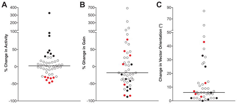
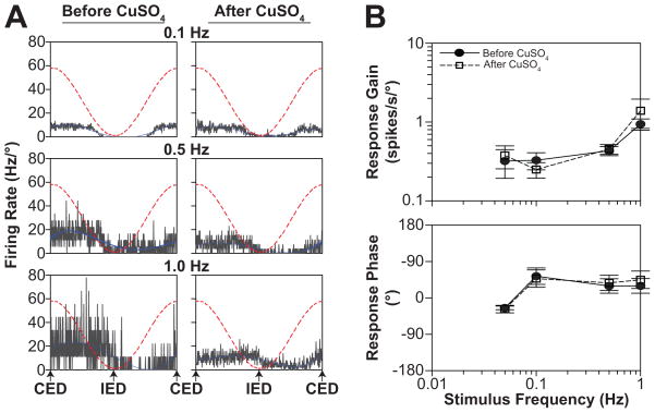
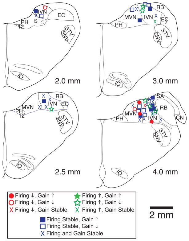
Neurons located in the caudal aspect of the vestibular nucleus complex have been shown to receive visceral inputs and project to brainstem regions that participate in generating emesis, such as nucleus tractus solitarius and the "vomiting region" in the lateral tegmental field (LTF). Consequently, it has been hypothesized that neurons in the caudal vestibular nuclei participate in triggering motion sickness and that visceral inputs to the vestibular nucleus complex can affect motion sickness susceptibility. To obtain supporting evidence for this hypothesis, we determined the effects of intragastric infusion of copper sulfate (CuSO4) on responses of neurons in the inferior and caudal medial vestibular nuclei to rotations in vertical planes. CuSO4 readily elicits nausea and emesis by activating gastrointestinal (GI) afferents. Infusion of CuSO4 produced a >30 % change in spontaneous firing rate of approximately one-third of neurons in the caudal aspect of the vestibular nucleus complex. These changes in firing rate developed over several minutes, presumably in tandem with the emetic response. The gains of responses to vertical vestibular stimulation of a larger fraction (approximately two-thirds) of caudal vestibular nucleus neurons were altered over 30 % by administration of CuSO4. The response gains of some units went up, and others went down, and there was no significant relationship with concurrent spontaneous firing rate change. These findings support the notion that the effects of visceral inputs on motion sickness susceptibility are mediated in part through the caudal vestibular nuclei. However, our previous studies showed that infusion of CuSO4 produced larger changes in response to vestibular stimulation of LTF neurons, as well as parabrachial nucleus neurons that are believed to participate in generating nausea. Thus, integrative effects of GI inputs on the processing of labyrinthine inputs must occur at brain sites that participate in eliciting motion sickness in addition to the caudal vestibular nuclei. It seems likely that the occurrence of motion sickness requires converging inputs to brain areas that generate nausea and vomiting from a variety of regions that process vestibular signals.


Figures

Figure 1
Figure 1
Changes in spontaneous firing rate of a vestibular nucleus neuron produced by the intragastric infusion of copper sulfate (CuSO4). A: a raster plot of neuronal activity before and following CuSO4 delivery. Each tick mark indicates the occurrence of an action potential. Shaded areas indicate periods of neuronal activity that are expanded in panels C–E. B: tracing of blood pressure recorded simultaneously with unit activity. C–E: 10 msec periods of unit activity recorded before (C), immediately after (D), and approximately 150 sec following (E) the infusion of CuSO4. Unit activity was sampled at 25,000 Hz. The spontaneous firing rate of the unit decreased after CuSO4 was provided.
Figure 2
Figure 2
A: average unit activity (top) and blood pressure (bottom) during 20-sec intervals before (gray bars) and after (white bars) the administration of CuSO4. Error bars indicate one SEM, and asterisks indicate intervals when unit activity and blood pressure were significantly and >30% different from those during the first 20 sec of recording (leftmost bar in each histogram). B: averaged responses to clockwise (CW) and counterclockwise (CCW) wobble stimuli (0.5 Hz, 5° amplitude) recorded before and after the delivery of CuSO4. Each panel shows unit activity, with a superimposed sine wave fit to the response. Responses to the following number of wobble rotations were averaged to generate each trace: pre-CuSO4 CW, 10; pre-CuSO4 CCW, 11; post-CuSO4 CW, 72; post-CuSO4 CCW, 43. Following the administration of CuSO4, the average response gain to wobble rotations decreased from 7.6 spikes/sec/° to 2.8 spikes/sec/°. Abbreviations: CED, contralateral ear down roll; IED, ipsilateral ear down roll; ND, nose-down pitch; NU, nose-up pitch.
Figure 3
Figure 3
Effects of intragastric CuSO4 on the spontaneous activity (A), gain of responses to wobble stimuli (B), and response vector orientation (C) of vestibular nucleus neurons. For A and B, the values plotted indicate: (pre-lesion value − post-lesion value)/pre-lesion value X 100%. Each symbol indicates values for a particular neuron; filled symbols denote data for the subset of units whose spontaneous activity changed >30% after CuSO4 was injected, with red symbols indicating the subset of units whose firing rate decreased. Horizontal lines indicate median values. Responses to wobble stimuli were so attenuated after CuSO4 in four cases that the signal-to-noise ratio no longer met our criteria for significance. Data for such cases were used to calculate the change in response gain (B), but not the change in response vector orientation (C). Hence, fewer data values are plotted in (C) than (A) or (B).
Figure 4
Figure 4
Effects of intragastric CuSO4 on the responses of vestibular nucleus neurons to fixed-plane tilts delivered near the plane of the response vector orientation. A: averaged responses of one neuron to roll tilts delivered at 0.1 Hz (5°), 0.5 Hz (5°) and 1 Hz (2.5°). Dashed red lines indicate table position. Following the administration of CuSO4, the response gain decreased 30% at 0.1 Hz, 45% at 0.5 Hz, and 32% at 1.0 Hz. However, response phases were similar before and after the injection of CuSO4. B: average Bode plots for all neurons whose responses to rotations in a single vertical plane were recorded before and after CuSO4 infusion. Response gains and phases were plotted with respect to stimulus position. Error bars indicate one SEM. Abbreviations: CED, contralateral ear down roll; IED, ipsilateral ear down roll.
Figure 5
Figure 5
Locations of neurons tested for the effects of CuSO4 on responses to whole-body tilts. Unit locations are plotted on standard transverse sections through the caudal medulla, which were generated through reference to Berman’s atlas (Berman 1968). Numbers below each section designate distance in mm rostral to the obex. Red symbols indicate units whose spontaneous firing rate decreased >30% following CuSO4, green symbols indicate units whose spontaneous firing rate increased >30%, and blue symbols indicate units whose spontaneous firing rate remained stable. Filled symbols indicate cells whose response gains to rotations increased >30% after CuSO4, open symbols indicate cells whose response gains decreased >30%, and “X”s indicate units whose response gains remained stable. Abbreviations: 12, hypoglossal nucleus; CN, cochlear nucleus; EC, external cuneate; IO, inferior olivary nucleus; PH, prepositus hypoglossi; IVN, inferior vestibular nucleus; MVN, medial vestibular nucleus; RB, restiform body; S, solitary nucleus; SA, stria acoustica; SNV, spinal trigeminal nucleus; STV, spinal trigeminal tract.

References

    1. Anderson JH, Blanks RHI, Precht W. Response characteristics of semicircular canal and otolith systems in the cat. I. Dynamic responses of primary vestibular fibers. Exp Brain Res. 1978;32:491–507. - PubMed
    1. Andrezik JA, Dormer KJ, Foreman RD, Person RJ. Fastigial nucleus projections to the brain stem in beagles: pathways for autonomic regulation. Neurosci. 1984;11:497–507. - PubMed
    1. Angaut P, Brodal A. The projection of the “vestibulocerebellum” onto the vestibular nuclei in the cat. Arch Ital Biol. 1967;105:441–479. - PubMed
    1. Ariumi H, Saito R, Nago S, Hyakusoku M, Takano Y, Kamiya H. The role of tachykinin NK-1 receptors in the area postrema of ferrets in emesis. Neurosci Lett. 2000;286:123–126. - PubMed
    1. Baker J, Goldberg J, Hermann G, Peterson B. Spatial and temporal response properties of secondary neurons that receive convergent input in vestibular nuclei of alert cats. Brain Res. 1984;294:138–143. - PubMed

Publication types