. 2013 Sep 1;591(17):4149-66.
doi: 10.1113/jphysiol.2013.255836. Epub 2013 May 27.

Stabilization of Kv4 protein by the accessory K(+) channel interacting protein 2 (KChIP2) subunit is required for the generation of native myocardial fast transient outward K(+) currents

Affiliations

Stabilization of Kv4 protein by the accessory K(+) channel interacting protein 2 (KChIP2) subunit is required for the generation of native myocardial fast transient outward K(+) currents

Nicholas C Foeger et al. J Physiol. .

Abstract

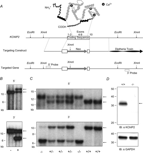
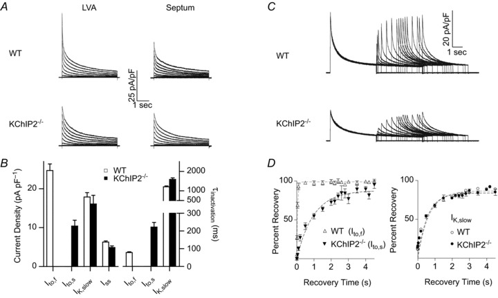
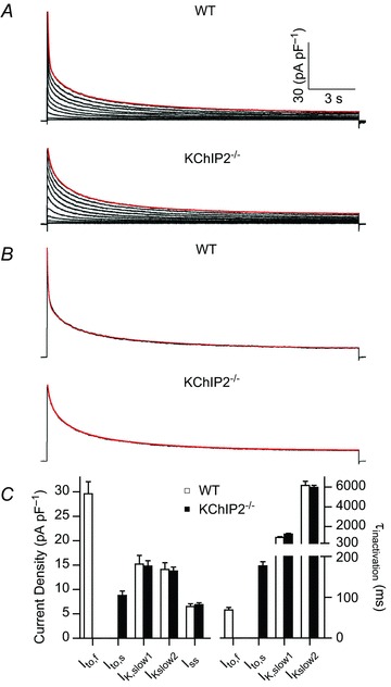
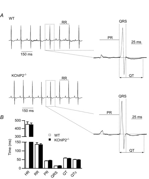
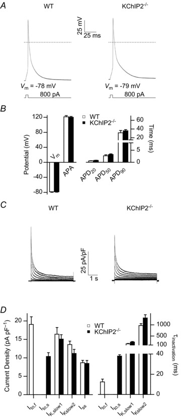
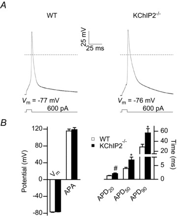
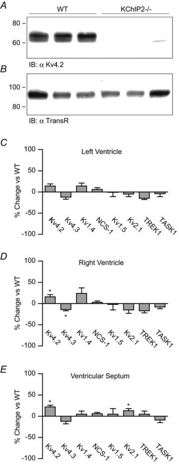
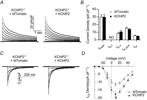
The fast transient outward K(+) current (Ito,f) underlies the early phase of myocardial action potential repolarization, contributing importantly to the coordinated propagation of activity in the heart and to the generation of normal cardiac rhythms. Native Ito,f channels reflect the tetrameric assembly of Kv4 pore-forming (α) subunits, and previous studies suggest roles for accessory and regulatory proteins in controlling the cell surface expression and the biophysical properties of Kv4-encoded Ito,f channels. Here, we demonstrate that the targeted deletion of the cytosolic accessory subunit, K(+) channel interacting protein 2 (KChIP2), results in the complete loss of the Kv4.2 protein, the α subunit critical for the generation of mouse ventricular Ito,f. Expression of the Kcnd2 (Kv4.2) transcript in KChIP2(-/-) ventricles, however, is unaffected. The loss of the Kv4.2 protein results in the elimination of Ito,f in KChIP2(-/-) ventricular myocytes. In parallel with the elimination of Ito,f, the slow transient outward K(+) current (Ito,s) is upregulated and voltage-gated Ca(2+) currents (ICa,L) are decreased. In addition, surface electrocardiograms and ventricular action potential waveforms in KChIP2(-/-) and wild-type mice are not significantly different, suggesting that the upregulation of Ito,s and the reduction in ICa,L compensate for the loss of Ito,f. Additional experiments revealed that Ito,f is not 'rescued' by adenovirus-mediated expression of KChIP2 in KChIP2(-/-) myocytes, although ICa,L densities are increased. Taken together, these results demonstrate that association with KChIP2 early in the biosynthetic pathway and KChIP2-mediated stabilization of Kv4 protein are critical determinants of native cardiac Ito,f channel expression.


Figures

Figure 1
Figure 1. Kcnip2 targeting construct and the generation of KChIP2−/− mice
A, the top schematic illustrates the general topography of KChIP2 and the approximate locations of Ca2+ binding (to the EF-hand motifs). Domains targeted for deletion are in grey and the physical maps of mouse Kcnip2, the targeting construct and the targeted allele are also illustrated. The regions selected for the generation of the 5′ and 3′ probes for Southern blot analysis are also indicated. B, XmnI-digested genomic DNA from transfected ES cells that survived the selection procedure were screened using the 5′ and 3′ probes; results from a negative (left lane) and a positive (right lane) clone are depicted. C, Southern blots of genomic tail DNA from WT (KChIP2+/+), homozygous targeted deletion (KChIP2−/−) and heterozygous (KChIP2+/−) animals probed with the 5′ and 3′ probes; the WT (5′: 11.7 kb; 3′: 11.2 kb) and targeted (5′: 13.9 kb; 3′: 6.3 kb) alleles are indicated by arrows. D, Western blots of brain lysates probed with a mouse monoclonal anti-KChIP2 antibody confirmed that no KChIP2 protein (arrow) is detected in KChIP2−/− animals.
Figure 2
Figure 2. The rapidly inactivating and recovering Ito,f is eliminated and electrical remodelling is evident in KChIP2−/− ventricular myocytes
A, representative whole-cell Kv currents, recorded at room temperature from WT and KChIP2−/− left ventricular apex (LVA) and interventricular septum (septum) myocytes in response to 4.5 s depolarizing voltage steps to test potentials between −60 and +40 mV (10 mV increments) from a holding potential of −70 mV; recorded currents were normalized for differences in cell size (whole-cell membrane capacitance) and current densities are plotted. As is evident, the rapid component of current decay, which is prominent in WT LVA cells, is absent in KChIP2−/− LVA myocytes, and the waveforms of the currents in the KChIP2−/− LVA and septum cells are quite similar. The decay phases of the outward Kv currents in WT and KChIP2−/− LVA cells were fitted to the sum of two exponentials and the mean current densities and Ito,f (Ito,s) and IK,slow inactivation time constants, determined from these fits, in WT (n= 37) and KChIP2−/− (n= 12) LVA myocytes, are presented in B. Similar analyses were completed to determine the amplitudes/densities and inactivation kinetics of the Kv currents in septum cells (see text). To examine the kinetics of Kv current recovery from inactivation, a three-pulse protocol was used: after inactivating the currents during 4.5 s pre-pulses to +30 mV, LVA cells were hyperpolarized to −70 mV for varying times (5 ms to 4.5 s) before test depolarizations to +30 mV (C). Representative current waveforms, recorded using this protocol from WT and KChIP2−/− LVA cells, are illustrated. As is evident, no rapidly recovering current is present in KChIP2−/− LVA cells. The amplitudes of Ito,f and IK,slow in WT LVA cells and the amplitudes of Ito,s and IK,slow in KChIP2−/− LVA cells (at +30 mV) after each recovery period were determined from double exponential fits to the decay phases of the currents (see Methods). These values were normalized to the current amplitudes in the same cell evoked after the 10 s recovery period and mean normalized recovery data for Ito,f▵ in WT LVA cells and Ito,s (▾), in KChIP2−/− LVA cells and for IK,slow (○,•) in WT (n= 8) and KChIP2−/− (n= 11) LVA cells are plotted as a function of recovery time in D; the recovery data for each of the currents are well described by single exponentials (dotted lines). The time constants (τrec) for Ito,f and Ito,s recovery determined from these fits in WT and KChIP2−/− LVA myocytes are 51 ± 1 and 1213 ± 285 ms, respectively. The τrec determined for IK,slow in KChIP2−/− (385 ± 17 ms) and WT (415 ± 21 ms) LVA myocytes are not significantly different.
Figure 3
Figure 3. Neither component of IK,slow, IK,slow1 or IK,slow2, is measurably altered in KChIP2−/− ventricular myocytes
A, representative whole-cell Kv currents, recorded at room temperature from WT and KChIP2−/− LVA myocytes in response to 20 s depolarizing voltage steps to test potentials between −60 and +60 mV (in 10 mV increments) from a holding potential of −70 mV; the currents were normalized to the whole cell membrane capacitance (in the same cell) and current densities are plotted. Outward currents (from the two representative cells in A) evoked at +60 mV are replotted in B and three-exponential fits to the decay phases of the currents are plotted as lines (in red) superimposed on the experimental data plotted as points (in black). The mean densities of Ito,f (Ito,s), IK,slow1, IK,slow2 and Iss, and the mean time constants of Ito,f (Ito,s), IK,slow1 and IK,slow2 inactivation, determined from these fits, in WT (n= 8) and KChIP2−/− (n= 19) LVA myocytes are presented in C. As is evident, the densities and the time constants of inactivation of IK,slow1 and IK,slow2 in WT and KChIP2−/− LVA myocytes are not significantly different (see text).
Figure 4
Figure 4. ECG waveforms are unaffected by the loss of KChIP2
A, representative telemetric ECG traces recorded from WT (n= 8) and KChIP2−/− (n= 7) animals are similar; no differences in heart rates or in the morphologies of the P waves, QRS complexes or T waves are evident. B, statistical analyses revealed no significant differences in the mean durations of the RR intervals, PR intervals, QRS complexes and QT intervals in WT and KChIP2−/− animals.
Figure 5
Figure 5. Action potential and Kv current waveforms recorded at physiological temperatures are similar in KChIP2−/− and WT LVA myocytes
A, representative action potentials, evoked in response to brief depolarizing current injections, in WT and KChIP2−/− LVA myocytes at 35–37°C are similar. B, mean resting membrane potentials (Vm), action potential amplitudes (APA) and action potential durations at 20% (APD20), 50% (APD50) and 90% (APD90) repolarization are not significantly different in WT (n= 13) and KChIP2−/− (n= 14) LVA myocytes. C, representative whole-cell Kv currents, recorded at physiological temperature (35–37°C), from WT and KChIP2−/− LVA myocytes in response to 4.5 s depolarizing voltage steps to test potentials between −60 and +40 mV (in 10 mV increments) from a holding potential of −70 mV; recorded currents were normalized to the whole-cell membrane capacitance (in the same cell) and current densities are plotted. The decay phases of the currents were fitted to the sum of three exponentials to provide the amplitudes and the time constants of inactivation of the individual current components. D, mean densities of Ito,f (Ito,s), IK,slow1, IK,slow2 and Iss, as well as the mean time constants of Ito,f (Ito,s), IK,slow1 and IK,slow2 inactivation, derived from these fits in WT (n= 14) and KChIP2−/− (n= 11) LVA myocytes.
Figure 6
Figure 6. Action potentials recorded at room temperatures are prolonged in KChIP2−/−, compared with WT, ventricular myocytes
A, representative action potentials, recorded at room temperature (22–23°C), in WT and KChIP2−/− LVA myocytes in response to brief depolarizing current injections. B, mean Vm and APA measured in KChIP2−/− (n= 14) and WT (n= 14) LVA cells at room temperature are not significantly different. In contrast to recordings obtained at physiological temperature (Fig. 5), however, mean APD20 (#P < 0.01), APD50 (*P < 0.05) and APD90 (*P < 0.05) values are significantly longer in recordings from KChIP2−/−, compared with WT, LVA myocytes at room temperature.
Figure 7
Figure 7. Kv4.2 protein is undetectable in KChIP2−/− ventricles
A, Western blots of membrane proteins prepared from WT and KChIP2−/− ventricles, probed with a rabbit polyclonal anti-Kv4.2 antibody, revealed that the Kv4.2 protein is undetectable in KChIP2−/− ventricles. The blots were also probed with a mouse monoclonal anti-transferrin receptor antibody to ensure equal protein loading of the samples (B). The expression levels of the transcripts encoding multiple Kv channel subunits in adult (8–14 weeks) WT (n= 6) and KChIP2−/− (n= 6) left ventricles (C), right ventricles (D) and interventricular septa (E) were examined using SYBR green quantitative RT-PCR. Mean percentage changes in the relative expression levels of each transcript in KChIP2−/−, compared with WT, left ventricles (C), right ventricles (D) and septa (E) are plotted. As is evident, in contrast to the Kv4.2 protein, the Kcnd2 transcript is not eliminated in KChIP2−/− ventricles. Indeed, the Kcnd2 transcript was modestly, but significantly (*P < 0.05), increased in the KChIP2−/− right ventricular and interventricular septum samples. The expression levels of the transcripts encoding the other Kvα subunits in KChIP2−/− ventricles were also similar to WT levels. There was a modest (∼10%) increase in the Kcnb1 (*P < 0.05) transcript in KChIP2−/− interventricular septa and the Kcnd3 transcript was modestly (*P < 0.05) decreased in KChIP2−/− right ventricles.
Figure 8
Figure 8. Increased expression of KChIP2 does not rescue Ito,f in KChIP2−/− myocytes, but does augment L-type Ca2+ current densities
Isolated KChIP2−/− ventricular myocytes were infected with an adenoviral construct encoding either tdTomato alone or tdTomato with KChIP2 (see Methods). Whole-cell Kv currents were recorded 36–48 h later from tdTomato-expressing cells as described in the legend to Fig. 2; currents were normalized to the whole-cell membrane capacitance (in the same cell) and current densities are plotted. A, Kv current waveforms in KChIP2−/− cells expressing tdTomato alone and tdTomato with KChIP2 are indistinguishable. B, mean peak Kv current, Ito,s, IK,slow and Iss densities in tdTomato- (n= 7) and tdTomato + KChIP2- (n= 4) expressing KChIP2−/− cells are not significantly different. C, whole-cell Cav currents, evoked in response to 400 ms depolarizing voltage steps to −40 to +50 mV (10 mV increments) from a holding potential of −70 mV, were also recorded from KChIP2−/− myocytes 36–48 h after infection with the tdTomato or tdTomato + KChIP2 adenovirus; recorded currents were normalized to whole-cell membrane capacitance (in the same cell), and current densities are plotted. D, mean Cav current densities are significantly (#P < 0.01) higher in tdTomato + KChIP2-expressing KChIP2−/− cells (n= 13) than in tdTomato-expressing KChIP2−/− cells (n= 14).

Similar articles

Cited by

References

    1. Aimond F, Kwak SP, Rhodes KJ, Nerbonne JM. Accessory Kvβ1 subunits differentially modulate the functional expression of voltage-gated K+ channels in mouse ventricular myocytes. Circ Res. 2005;96:451–458. - PubMed
    1. An WF, Bowlby MR, Betty M, Cao J, Ling HP, Mendoza G, Hinson JW, Mattsson KI, Strassle BW, Trimmer JS, Rhodes KJ. Modulation of A-type potassium channels by a family of calcium sensors. Nature. 2000;403:553–556. - PubMed
    1. Antzelevitch C, Sicouri S, Litovsky S, Lukas A, Krishnan S, Di Diego J, Gintant G, Liu D. Heterogeneity within the ventricular wall. Electrophysiology and pharmacology of epicardial, endocardial, and M cells. Circ Res. 1991;69:1427–1449. - PubMed
    1. Bähring R, Dannenberg J, Peters HC, Leicher T, Pongs O, Isbrandt D. Conserved Kv4 N-terminal domain critical for effects of Kv channel-interacting protein 2.2 on channel expression and gating. J Biol Chem. 2001;276:23888–23894. - PubMed
    1. Barry DM, Xu H, Schuessler RB, Nerbonne JM. Functional knockout of the transient outward current, long-QT syndrome, and cardiac remodeling in mice expressing a dominant-negative Kv4 alpha subunit. Circ Res. 1998;83:560–567. - PubMed

Publication types