Review
. 2013 Jun 25;5(3):167-81.
doi: 10.1042/AN20120095.

LGI proteins in the nervous system

Affiliations
Review

LGI proteins in the nervous system

Linde Kegel et al. ASN Neuro. .

Abstract

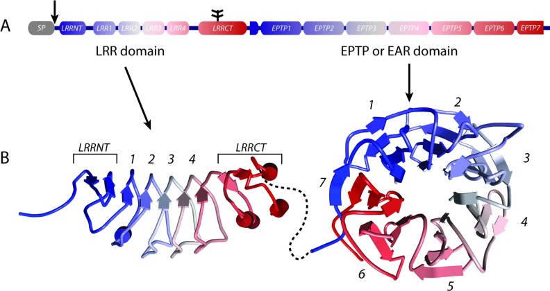
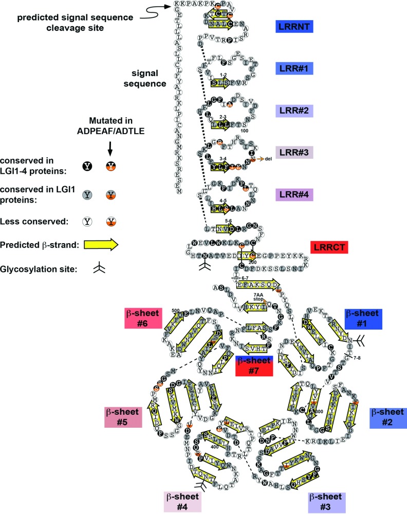
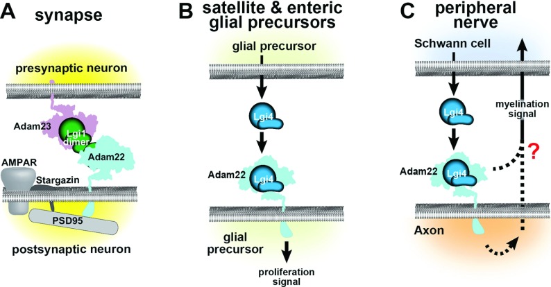
The development and function of the vertebrate nervous system depend on specific interactions between different cell types. Two examples of such interactions are synaptic transmission and myelination. LGI1-4 (leucine-rich glioma inactivated proteins) play important roles in these processes. They are secreted proteins consisting of an LRR (leucine-rich repeat) domain and a so-called epilepsy-associated or EPTP (epitempin) domain. Both domains are thought to function in protein-protein interactions. The first LGI gene to be identified, LGI1, was found at a chromosomal translocation breakpoint in a glioma cell line. It was subsequently found mutated in ADLTE (autosomal dominant lateral temporal (lobe) epilepsy) also referred to as ADPEAF (autosomal dominant partial epilepsy with auditory features). LGI1 protein appears to act at synapses and antibodies against LGI1 may cause the autoimmune disorder limbic encephalitis. A similar function in synaptic remodelling has been suggested for LGI2, which is mutated in canine Benign Familial Juvenile Epilepsy. LGI4 is required for proliferation of glia in the peripheral nervous system and binds to a neuronal receptor, ADAM22, to foster ensheathment and myelination of axons by Schwann cells. Thus, LGI proteins play crucial roles in nervous system development and function and their study is highly important, both to understand their biological functions and for their therapeutic potential. Here, we review our current knowledge about this important family of proteins, and the progress made towards understanding their functions.


Figures

Figure 1
Figure 1. Structural characteristics of the LGI protein family
In (A), the general domain structure of the LGI protein family is depicted. All LGI proteins have an SP (signal peptide) that is cleaved off (arrow in A) and is not included in the putative protein structures shown in (B). The glycosylation site present in all LGI members is indicated in (A) with a branched line structure. The putative structures of the LRR domain and EPTP domain were predicted separately using the HHpred tool (http://toolkit.tuebingen.mpg.de/hhpred) and the WDR5 protein structure (PDB 2GNQA) as template. Structures were visualized using the Accelrys Discovery Studio visualizer. The structure is colour-coded from N-terminus (blue) to C-terminus (red) and corresponds with the colour code in (A). The colour-code graphically reveals the Velcro β-strand (blue) interacting with the last β-strand (red) of the seventh EPTP module to zip up the EPTP domain structure. How the LRR domain and the EPTP domain are oriented towards each other is unknown. The stippled black line does not represent any structural feature but is only intended to show the linkage between the two domains.
Figure 2
Figure 2. A schematic diagram of human LGI1 protein
In this ‘exploded’ view of LGI1, the individual LRR and EPTP modules are separated from one another; dashed lines connect amino acids that are linked in the intact protein. Amino acids are represented as filled, shaded or open circles, depending on their level of conservation. The bottom half of circles for amino acids that are changed by point mutations are coloured orange; all of these mutations are from human except for the mutation at Leu385, which is from rat. For simplicity, frameshift mutations are omitted, with the exception of one at the C-terminus that provides evidence that the ‘Velcro’ model of β-propeller closure, in which the N-terminal β-strand is included in the seventh EPTP propeller fold. Human LGI1 mutations were obtained from (de Bellescize et al., ; Nobile et al., ; Di Bonaventura et al., ; Ho et al., 2012) and the rat mutation from (Baulac et al., 2012). For the EPTP domain, the β-propeller blades, which in large part would intersect the plane of the figure in the intact protein, have been laid flat. β-strands are outlined by yellow arrows (Leonardi et al., 2011). Disulfide bonds are depicted as solid lines, and glycosylation sites have a branched line structure. Boundaries of the eight LGI1 exons are presented as lines through the sequence, with the relevant exon numbers juxtaposed. Every 100th amino acid is labelled.
Figure 3
Figure 3. Possible mechanisms by which LGI proteins participate in cell–cell interactions
Panels (A–C) depict several possible mechanisms by which LGI1 controls synapse development and function, and LGI4 controls PNS development. Note that these mechanisms are not mutually exclusive; the figure simply summarizes what currently is known or postulated for each interaction. (A) At synapses, LGI1 dimers may form a bridge between post-synaptic ADAM22, which is linked to the stargazin/AMPAR complex through PSD95 and stargazin, and presynaptic ADAM23 (Fukata et al., 2010). LGI1 has an effect on Kv1.1 inactivation through mechanisms that are unclear (Fukata et al., 2006). (B) In peripheral glial precursors, LGI4 appears to act as a paracrine or autocrine factor through binding to ADAM22 (Nishino et al., 2010). (C) During Schwann cell ensheathment and myelination of axons, LGI4 binds to the disintegrin domain of ADAM22. This interaction could trigger signalling or protein localization within the axon, thereby activating signal(s) to the Schwann cell. An LGI4–ADAM22–PSD95 interaction could cluster proteins at the axonal membrane. Alternatively, LGI4 binding to ADAM22 could induce the formation of protein complex(es) extracellularly. LGI4 might modulate ADAM22–integrin interactions, which also utilize the ADAM22 disintegrin domain (D’Abaco et al., 2006).

References

    1. Amemiya CT, Alföldi J, Lee AP, Fan S, Philippe H, Maccallum I, Braasch I, Manousaki T, Schneider I, Rohner N, et al. The African coelacanth genome provides insights into tetrapod evolution. Nature. 2013;496:311–316. - PMC - PubMed
    1. Barth AL, Malenka RC. NMDAR EPSC kinetics do not regulate the critical period for LTP at thalamocortical synapses. Nat Neurosci. 2001;4:235–236. - PubMed
    1. Baulac S, Ishida S, Mashimo T, Boillot M, Fumoto N, Kuwamura M, Ohno Y, Takizawa A, Aoto T, Ueda M, Ikeda A, LeGuern E, Takahashi R, Serikawa T. A rat model for LGI1-related epilepsies. Human molecular genetics. 2012;21:3546–3557. - PubMed
    1. Berghuis B, Brilstra EH, Lindhout D, Baulac S, de Haan GJ, van Kempen M. Hyperactive behavior in a family with autosomal dominant lateral temporal lobe epilepsy caused by a mutation in the LGI1/epitempin gene. Epilepsy & behavior: E&B. 2013;28:41–46. - PubMed
    1. Bermingham JR, Jr, Scherer SS, O’Connell S, Arroyo E, Kalla KA, Powell FL, Rosenfeld MG. Tst-1/Oct-6/SCIP regulates a unique step in peripheral myelination and is required for normal respiration. Genes Dev. 1996;10:1751–1762. - PubMed

Publication types

MeSH terms

Supplementary concepts

LinkOut - more resources