Review
. 2013 Jun;27(3):381-409.
doi: 10.1016/j.hoc.2013.02.002. Epub 2013 Apr 11.

Harnessing the platelet signaling network to produce an optimal hemostatic response

Affiliations
Review

Harnessing the platelet signaling network to produce an optimal hemostatic response

Lawrence F Brass et al. Hematol Oncol Clin North Am. 2013 Jun.

Abstract

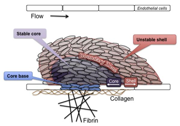
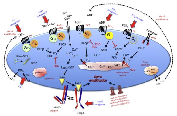
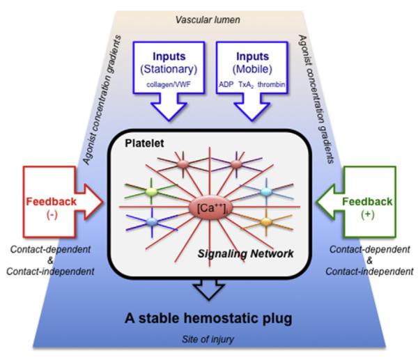
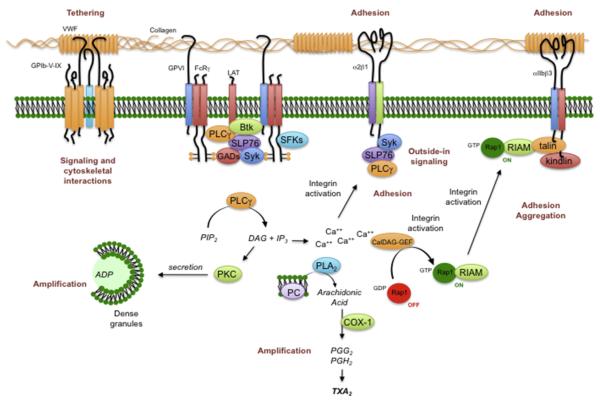
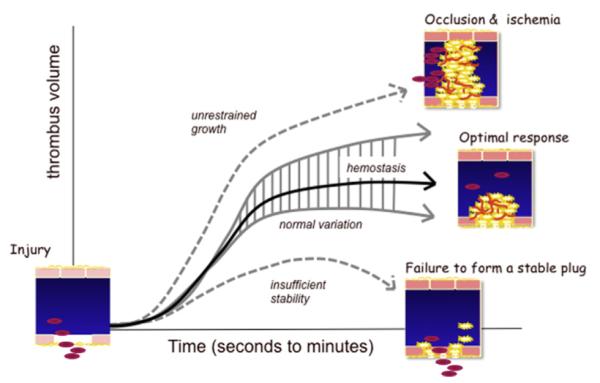
Once released into the circulation by megakaryocytes, circulating platelets can undergo rapid activation at sites of vascular injury and resist unwarranted activation, which can lead to heart attacks and strokes. Historically, the signaling mechanisms underlying the regulation of platelet activation have been approached as a collection of individual pathways unique to agonist. This review takes a different approach, casting platelet activation as the product of a signaling network, in which activating and restraining mechanisms interact in a flexible network that regulates platelet adhesiveness, cohesion between platelets, granule secretion, and the formation of a stable hemostatic thrombus.


Figures

Fig. 1
Fig. 1
Structural heterogeneity in the hemostatic plug. Recent observations of the platelet response to acute vascular injury in vivo show that there are distinct regions within the hemostatic thrombus where platelets are either more or less activated and where fibrin tends to accumulate. A stable core of closely packed, fully activated platelets is overlaid by an outer shell of less-stable, less-activated platelets, with a boundary zone in between. Fibrin is found within the thrombus core and in a fibrin network formed from fibrinogen that escapes before hemostasis can be achieved.
Fig. 2
Fig. 2
An overview of some of the major signaling pathways of platelet activation. Targets for antiplatelet agents that are in clinical use or in clinical trials are indicated in blue. Inhibitors are in red. AA, arachidonic acid; IP3, inositol-1,4,5-trisphosphate; PAR, protease-activated receptor; PGI2, prostaglandin I2; PKA, protein kinase A; PKC, protein kinase C; PLC, phospholipase C; RGS, regulator of G protein signaling; TXA2, thromboxane A2.
Fig. 3
Fig. 3
A network-centric view of platelet activation. Signaling in platelets is represented as a balanced network, in which the effects of multiple inputs (mainly agonists) are modulated by positive and negative feedback. Signaling pathways communicate with each other and share nodal points in the network. The extent of activation of any given platelet varies according to the combination and concentration of the agonists to which the platelet is exposed and the sum of the feedback to which it is subjected. Gradients of agonist concentration are expected to be highest at the vessel wall near the site of injury (bottom) and lowest in the thrombus shell (top). Referring to the model in Fig. 1, platelets in the core of the thrombus are most subject to thrombin, collagen, and contact-dependent feedback, whereas those in the shell are affected primarily by soluble mediators such as ADP and TxA2.
Fig. 4
Fig. 4
Platelet activation by collagen. Platelets use several different molecular complexes to support platelet activation by collagen. These complexes include (1) VWF-mediated binding of collagen to the GPIb-IX-V complex and integrin αIIbβ3 and (2) a direct interaction between collagen and both the integrin α2β1 and the GPVI/FcRγ-chain complex. Clustering of GPVI results in the phosphorylation of tyrosine residues in the FcRγ cytoplasmic domain, followed by the activation of the tyrosine kinase, Syk. One consequence of Syk activation is the activation of PLCγ2, leading to phosphoinositide hydrolysis, secretion of ADP, and the production of TXA2. COX-1, cyclooxygenase 1; DAG, diacylglycerol; PG, prostaglandin; PI3K, phosphatidylinositol 3-kinase; PKC, protein kinase C; PLA2, phospholipase A2.
Fig. 5
Fig. 5
Formation of an optimal platelet plug. Vascular injury produces a hemostatic response that can be too aggressive (leading to occlusion and ischemia), inadequate (leading to further bleeding), or optimal. This model suggests that an optimal response varies in detail (hence, a range of normal), but is best viewed as a response that results in hemostasis with a minimum of blood loss and an avoidance of unwarranted vascular occlusion. In the setting of a vascular wall disease such as atherosclerosis, the rapid accumulation of platelets on top of a ruptured plaque represents an escape from normal restraints.

References

    1. Kanaji S, Fahs SA, Shi Q, et al. Contribution of platelet versus endothelial VWF to platelet adhesion and hemostasis. J Thromb Haemost. 2012;10(8):1646–52. - PMC - PubMed
    1. Reininger AJ, Heijnen HF, Schumann H, et al. Mechanism of platelet adhesion to von Willebrand factor and microparticle formation under high shear stress. Blood. 2006;107:3537–45. - PMC - PubMed
    1. Ruggeri ZM, Orje JN, Habermann R, et al. Activation-independent platelet adhesion and aggregation under elevated shear stress. Blood. 2006;108:1903–10. - PMC - PubMed
    1. Munnix IC, Cosemans JM, Auger JM, et al. Platelet response heterogeneity in thrombus formation. Thromb Haemost. 2009;102:1149–56. - PubMed
    1. Nesbitt WS, Westein E, Tovar-Lopez FJ, et al. A shear gradient-dependent platelet aggregation mechanism drives thrombus formation. Nat Med. 2009;15:665–73. - PubMed